

Children with back pain - a radiologist's approach
- PMID: 37701175
- PMCID: PMC10493862
- DOI: 10.5114/pjr.2023.130977
Children with back pain - a radiologist's approach
Abstract
Purpose: The aim of the study was to analyse magnetic resonance imaging (MRI) of paediatric patients referred because of back pain.
Material and methods: The retrospective analysis included the medical records of 328 patients referred in 2020-2022 to the Department of Paediatric Radiology for spine examination. The criterion for inclusion in the analysed group was back pain as the dominant symptom. This symptom occurred in 20% (68 patients) of referrals for MRI examinations. The examination was performed with the 3T Magnetom Spectra.
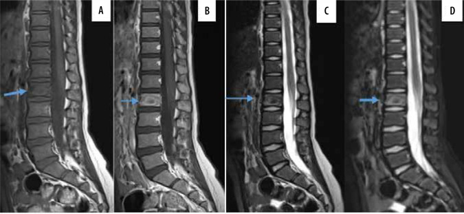
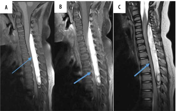
Results: In 68 patients aged 2 to 17 years, with back pain as the first diagnosis, 53% (36 patients - 16 girls and 20 boys) showed abnormalities. The rest of the tests were assessed as normal. Among the patients with an abnormal MR image, the largest group were children with degenerative changes diagnosed: 10 children (28%) aged 13-17 years. In 9 patients (25%) aged 2-16 years the final diagnosis qualified the patients to the group of oncological diagnoses. Another group of 7 (19%) patients, aged 6-14 years, comprised children diagnosed with inflammation. The group of 5 patients, aged 3-17 years, presented symptoms most likely related to the trauma. One 7-year-old boy was diagnosed with large calcifications within the intervertebral disc.
Conclusions: Back pain, with accompanying neurological symptoms, should not be underestimated. Although in most clinical situations the MR image is normal, in the case of persistent symptoms and neurological abnormalities confirmed by the clinician, extending the diagnostics with MR imaging should be considered. This imaging can accelerate the correct diagnostic path or make a very precise diagnosis.
Keywords: back pain; children; imaging; magnetic resonance.
© Pol J Radiol 2023.
Conflict of interest statement
The authors report no conflict of interest.
Figures


Similar articles
-
Seropositive Neuromyelitis Optica in a Case of Undiagnosed Ankylosing Spondylitis: A Neuro-Rheumatological Conundrum.Qatar Med J. 2022 Jul 7;2022(3):29. doi: 10.5339/qmj.2022.29. eCollection 2022. Qatar Med J. 2022. PMID: 35864917 Free PMC article.
-
Juvenile degenerative disc disease: a report of 76 cases identified by magnetic resonance imaging.Spine J. 2007 May-Jun;7(3):332-7. doi: 10.1016/j.spinee.2006.03.008. Epub 2007 Jan 24. Spine J. 2007. PMID: 17482117
-
When does a radiologist's recommendation for follow-up result in high-cost imaging?Radiology. 2012 Feb;262(2):544-9. doi: 10.1148/radiol.11111091. Epub 2011 Nov 14. Radiology. 2012. PMID: 22084210
-
The cost-effectiveness of magnetic resonance imaging for investigation of the knee joint.Health Technol Assess. 2001;5(27):1-95. doi: 10.3310/hta5270. Health Technol Assess. 2001. PMID: 11532240 Review.
-
Lumbar Spondylodiscitis Mimicking Cholecystitis: A Case Report and Review of Literature.J Neurol Surg A Cent Eur Neurosurg. 2023 Jan;84(1):95-102. doi: 10.1055/a-1811-7393. Epub 2022 Mar 30. J Neurol Surg A Cent Eur Neurosurg. 2023. PMID: 35354214
Cited by
-
Clinical presentation and imaging findings in juvenile-onset back pain: a ten-year hospital-based retrospective analysis in Douala (Cameroon).Front Pediatr. 2024 Jul 2;12:1424391. doi: 10.3389/fped.2024.1424391. eCollection 2024. Front Pediatr. 2024. PMID: 39015207 Free PMC article.
References
-
- Achar S, Yamanaka J. Back pain in children and adolescents. Am Fam Physician 2020; 102: 19-28. - PubMed
-
- Lamb M, Brenner JS. Back pain in children and adolescents. Pediatr Rev 2020; 41: 557-569. - PubMed
-
- Roberts SB, Calligeros K, Tsirikos AI. Evaluation and management of paediatric and adolescent back pain: epidemiology, presentation, investigation, and clinical management: a narrative review. J Back Musculoskelet Rehabil 2019; 32: 955-988. - PubMed
-
- Dunn AJ, Campbell RS, Mayor PE, et al. . Radiological findings and healing patterns of incomplete stress fractures of the pars interarticularis. Skeletal Radiol 2008; 37: 443-450. - PubMed
LinkOut - more resources
Full Text Sources