. 2023 May 19;11(1):coad031.
doi: 10.1093/conphys/coad031. eCollection 2023.

Infectious agents and their physiological correlates in early marine Chinook salmon (Oncorhynchus tshawytscha)

Affiliations

Infectious agents and their physiological correlates in early marine Chinook salmon (Oncorhynchus tshawytscha)

Yuwei Wang et al. Conserv Physiol. .

Abstract

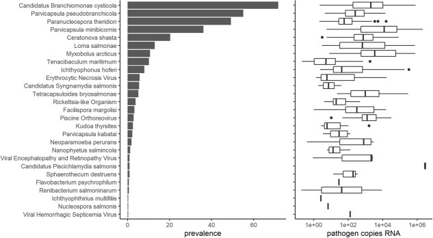
The early marine life of Pacific salmon is believed to be a critical period limiting population-level survival. Recent evidence suggests that some infectious agents are associated with survival but linkages with underlying physiological mechanisms are lacking. While challenge studies can demonstrate cause and effect relationships between infection and pathological change or mortality, in some cases pathological change may only manifest in the presence of environmental stressors; thus, it is important to gain context from field observations. Herein, we examined physiological correlates with infectious agent loads in Chinook salmon during their first ocean year. We measured physiology at the molecular (gene expression), metabolic (plasma chemistry) and cellular (histopathology) levels. Of 46 assayed infectious agents, 27 were detected, including viruses, bacteria and parasites. This exploratory study identified.

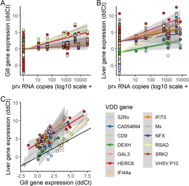
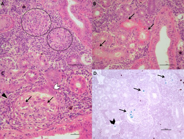
a strong molecular response to viral disease and pathological change consistent with jaundice/anemia associated with Piscine orthoreovirus,strong molecular signals of gill inflammation and immune response associated with gill agents `Candidatus Branchiomonas cysticola' and Parvicapsula pseudobranchicola,a general downregulation of gill immune response associated with Parvicapsula minibicornis complementary to that of P. pseudobranchicola.Importantly, our study provides the first evidence that the molecular activation of viral disease response and the lesions observed during the development of the PRV-related disease jaundice/anemia in farmed Chinook salmon are also observed in wild juvenile Chinook salmon.


Conflict of interest statement

The authors have no conflicts to declare.

Figures

Figure 1
Figure 1
Capture locations (points) of Chinook salmon sampled around Vancouver Island (center) by mid-water trawl from 2012 to 2014. Red rectangle in inset map indicates the extent of the main map. Point color and shape represents the capture regions assigned for statistical analysis (red circles = West Coast Vancouver Island, green triangles = Southeast VI, blue squares = Northeast VI). For each capture region, pie charts depict the population origin for the sampled fish (colors in legend correspond to pie charts only; ECVI = East Coast Vancouver Island, WCVI = West Coast Vancouver Island).
Figure 2
Figure 2
Correlations of gene expression (2−ΔΔCt) for individual genes with principal component 1 for each functional gene group. Gill is presented on left (blue) and liver is presented on right (orange). Osmoregulation gene expression was only analyzed for gill tissue.
Figure 3
Figure 3
Prevalence and load of all pathogens detected in Chinook salmon sampled around Vancouver Island, 2012–2014. Load data are presented on the log10 scale (only positive detections included in boxplot).
Figure 4
Figure 4
Heatmap of coefficients for each pathogen in models for blood plasma variables and the first principal components of functional gene groups in gill and liver tissue. Cell values represent the change in units of standard deviation for the plasma or gene variable associated with a standard deviation increase in log pathogen load (from mixed tissue). Two asterisks in a cell indicate an FDR-adjusted p value < 0.05 and a single asterisk indicates a p value < 0.05 prior to FDR adjustment.
Figure 5
Figure 5
The expression of genes associated with viral disease development (VDD) showed a positive correlation with piscine orthoreovirus load in gill (A, 9 positives in 292 samples) and liver (B, 9 positives in 263 samples) tissues of Chinook salmon captured during their first ocean year. Furthermore, VDD gene expression in gill and liver were well-correlated across individuals with PRV detections (C). Lines indicate a simple linear regression for each gene with accompanying 95% confidence interval (gray fill). Black line in C represents a 1:1 relationship.
Figure 6
Figure 6
Piscine orthoreovirus positive (PRV+) fish had lesions consistent with PRV-related jaundice/anemia (Fish B2159). A) Hematoxylin and eosin (H & E) staining shows focal endocarditis (dotted circles) in the spongy layer of the myocardium (scale bar = 200 μm). B) In situ hybridization of the same field reveals PRV in focal inflammatory infiltrates. C) Renal tubular vacuolar degeneration (arrows) leading to tubule necrosis (white arrowhead) in the kidney (scale bar = 50 μm). D) Blood congestion and hemosiderin deposits (arrows) in the spleen (scale bar = 1000 μm).
Figure 7
Figure 7
Epitheliocysts in the gill lamellae contained ‘Candidatus Branchiomonas cysticola’ (Fish B6930). A) Epitheliocysts in the gill lamellae (arrows) and associated proliferative/inflammatory reaction (asterisks, scale bar = 10 μm). B) Same field, using ISH to confirm that the epitheliocysts were infected with ‘Ca. B. cysticola’ (in red, scale bar = 10 μm).
Figure 8
Figure 8
Lesions consistent with Parvicapsula minibicornis, and the pathogen itself, were found in kidney tissues (Fish B5083). A) Two different degrees of Glomerulonephritis (dashed circle): the glomerulus on the right is in a more advanced stage of necrosis (moderate), while the one on the left still shows a few morphological features (mild) and generalized interstitial hyperplasia (scale bar = 50 μm) B) Eosinophilic lipoprotein droplets (arrows) were visible in some renal tubules (scale bar = 50 μm). C) Glomerulonephritis (triangle head), hypertrophy/hyperplasia of Bowman’s capsule (white arrowhead) and P. minibicornis pre-sporogonic forms (arrows) (scale bar = 20 μm). D) P. minibicornis (blue) detection by ISH on both glomeruli (arrows) and in the lumen of renal tubules (black arrowhead) (scale bar = 100 μm).

Similar articles

Cited by

References

    1. Akbarzadeh A, Günther OP, Houde AL, Li S, Ming TJ, Jeffries KM, Hinch SG, Miller KM (2018) Developing specific molecular biomarkers for thermal stress in salmonids. BMC Genomics 19: 749. 10.1186/s12864-018-5108-9. - DOI - PMC - PubMed
    1. Bakke TA, Harris PD (1998) Diseases and parasites in wild Atlantic salmon (Salmo salar) populations. Can J Fish Aquat Sci 55: 247–266. 10.1139/d98-021. - DOI
    1. Barrett DE, Bartholomew JL (2021) A tale of two fish: comparative transcriptomics of resistant and susceptible steelhead following exposure to Ceratonova shasta highlights differences in parasite recognition. PloS One 16: 1–31, e0234837. 10.1371/journal.pone.0234837. - DOI - PMC - PubMed
    1. Bartholomew JL, Atkinson SD, Hallett SL (2006) Involvement of manayunkia speciosa (annelida: polychaeta: sabellidae) in the life cycle of parvicapsula minibicornis, a myxozoan parasite of pacific salmon. J Parasitol 92: 742–748. 10.1645/GE-781R.1. - DOI - PubMed
    1. Bass AL, Hinch SG, Teffer AK, Patterson DA, Miller KM (2017) A survey of microparasites present in adult migrating Chinook salmon (Oncorhynchus tshawytscha) in South-Western British Columbia determined by high-throughput quantitative polymerase chain reaction. J Fish Dis 40: 453–477. 10.1111/jfd.12607. - DOI - PubMed