Capra cartilage-derived peptide delivery via carbon nano-dots for cartilage regeneration
- PMID: 37701494
- PMCID: PMC10493328
- DOI: 10.3389/fbioe.2023.1213932
Capra cartilage-derived peptide delivery via carbon nano-dots for cartilage regeneration
Abstract
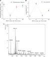
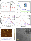
Targeted delivery of site-specific therapeutic agents is an effective strategy for osteoarthritis treatment. The lack of blood vessels in cartilage makes it difficult to deliver therapeutic agents like peptides to the defect area. Therefore, nucleus-targeting zwitterionic carbon nano-dots (CDs) have immense potential as a delivery vehicle for effective peptide delivery to the cytoplasm as well as nucleus. In the present study, nucleus-targeting zwitterionic CDs have been synthesized as delivery vehicle for peptides while also working as nano-agents towards optical monitoring of cartilage healing. The functional groups of zwitterion CDs were introduced by a single-step microwave assisted oxidation procedure followed by COL II peptide conjugation derived from Capra auricular cartilage through NHS/EDC coupling. The peptide-conjugated CDs (PCDs) allows cytoplasmic uptake within a short period of time (∼30 m) followed by translocation to nucleus after ∼24 h. Moreover, multicolor fluorescence of PCDs improves (blue, green, and read channel) its sensitivity as an optical code providing a compelling solution towards enhanced non-invasive tracking system with multifunctional properties. The PCDs-based delivery system developed in this study has exhibited superior ability to induce ex-vivo chondrogenic differentiation of ADMSCs as compared to bare CDs. For assessment of cartilage regeneration potential, pluronic F-127 based PCDs hydrogel was injected to rabbit auricular cartilage defects and potential healing was observed after 60 days. Therefore, the results confirm that PCDs could be an ideal alternate for multimodal therapeutic agents.
Keywords: carbon nano dots; cartilage regeneration; collagen II; peptide synthesis; zwitterion.
Copyright © 2023 Maity, Kapat, Poddar, Bora, Das, Das, Ganguly, Das, Dhara, Mandal, Roy Chowdhury, Mukherjee and Dhara.
Conflict of interest statement
The authors declare that the research was conducted in the absence of any commercial or financial relationships that could be construed as a potential conflict of interest.
Figures


Similar articles
-
Cell Nucleus-Targeting Zwitterionic Carbon Dots.Sci Rep. 2015 Dec 22;5:18807. doi: 10.1038/srep18807. Sci Rep. 2015. PMID: 26689549 Free PMC article.
-
Nano-carrier for gene delivery and bioimaging based on pentaetheylenehexamine modified carbon dots.J Colloid Interface Sci. 2023 Jun;639:180-192. doi: 10.1016/j.jcis.2023.02.046. Epub 2023 Feb 15. J Colloid Interface Sci. 2023. PMID: 36805743
-
Auricular cartilage regeneration using different types of mesenchymal stem cells in rabbits.Biol Res. 2022 Dec 26;55(1):40. doi: 10.1186/s40659-022-00408-z. Biol Res. 2022. PMID: 36572914 Free PMC article.
-
New insight into the engineering of green carbon dots: Possible applications in emerging cancer theranostics.Talanta. 2020 Mar 1;209:120547. doi: 10.1016/j.talanta.2019.120547. Epub 2019 Nov 13. Talanta. 2020. PMID: 31892009 Review.
-
Peptide-Conjugated Nano Delivery Systems for Therapy and Diagnosis of Cancer.Pharmaceutics. 2021 Sep 9;13(9):1433. doi: 10.3390/pharmaceutics13091433. Pharmaceutics. 2021. PMID: 34575511 Free PMC article. Review.
Cited by
-
Recent Uses of Lipid Nanoparticles, Cell-Penetrating and Bioactive Peptides for the Development of Brain-Targeted Nanomedicines against Neurodegenerative Disorders.Nanomaterials (Basel). 2023 Nov 23;13(23):3004. doi: 10.3390/nano13233004. Nanomaterials (Basel). 2023. PMID: 38063700 Free PMC article. Review.
-
Fabrication and Applications of Magnetic Polymer Composites for Soft Robotics.Micromachines (Basel). 2023 Nov 29;14(12):2173. doi: 10.3390/mi14122173. Micromachines (Basel). 2023. PMID: 38138344 Free PMC article. Review.
-
Carbon dots-based drug delivery for bone regeneration.Front Bioeng Biotechnol. 2025 May 29;13:1613901. doi: 10.3389/fbioe.2025.1613901. eCollection 2025. Front Bioeng Biotechnol. 2025. PMID: 40511299 Free PMC article. Review.
References
-
- Aleksandra Loczechin (2019). Carbon nanomaterials as antibacterial and antiviral alternatives. Other. Université de Lille; Ruhr-Universität. Available at: https://theses.hal.science/tel-03622608 .
LinkOut - more resources
Full Text Sources

