. 2023 Aug 25;26(10):107715.
doi: 10.1016/j.isci.2023.107715. eCollection 2023 Oct 20.

Active shrinkage protects neurons following axonal transection

Affiliations

Active shrinkage protects neurons following axonal transection

Mehmet Şerif Aydın et al. iScience. .

Abstract

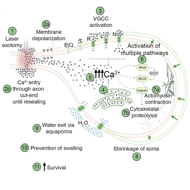
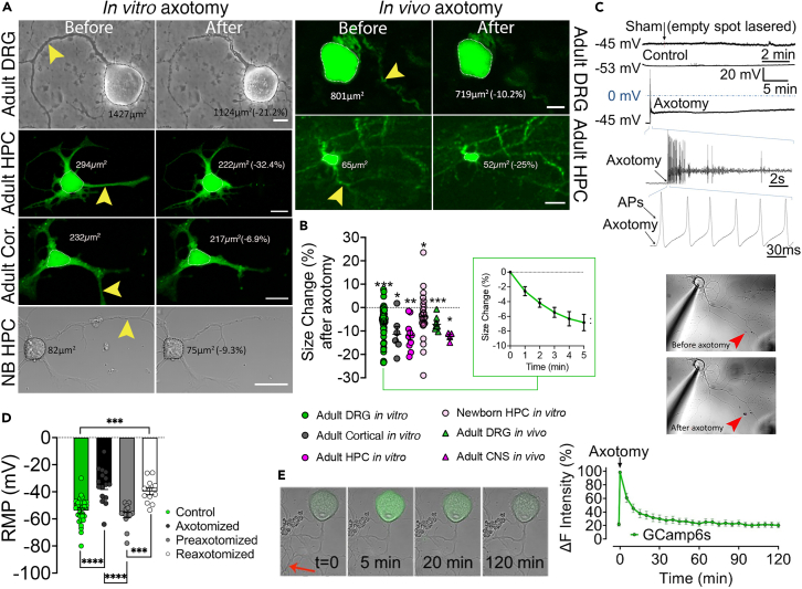
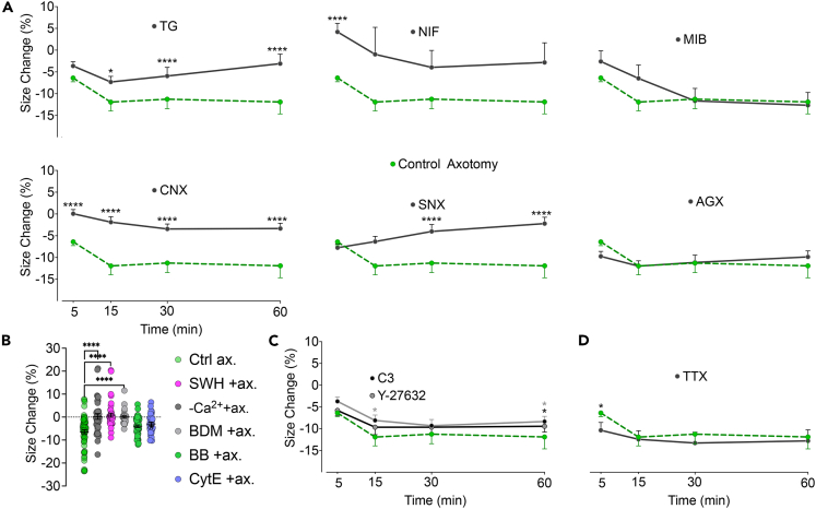
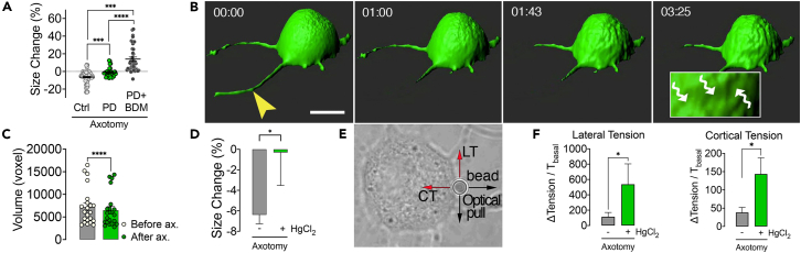
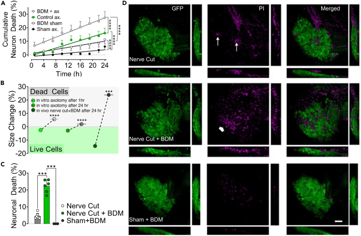
Trauma, vascular events, or neurodegenerative processes can lead to axonal injury and eventual transection (axotomy). Neurons can survive axotomy, yet the underlying mechanisms are not fully understood. Excessive water entry into injured neurons poses a particular risk due to swelling and subsequent death. Using in vitro and in vivo neurotrauma model systems based on laser transection and surgical nerve cut, we demonstrated that axotomy triggers actomyosin contraction coupled with calpain activity. As a consequence, neurons shrink acutely to force water out through aquaporin channels preventing swelling and bursting. Inhibiting shrinkage increased the probability of neuronal cell death by about 3-fold. These studies reveal a previously unrecognized cytoprotective response mechanism to neurotrauma and offer a fresh perspective on pathophysiological processes in the nervous system.

Keywords: Biological sciences; Neuroscience; Physiology.


Conflict of interest statement

Authors declare that they have no competing interests.

Figures

None
Graphical abstract
Figure 1
Figure 1
Axotomy causes a reduction in cell size, which is preceded by calcium influx and membrane depolarization (A) Images show adult mouse dorsal root ganglion (DRG), hippocampal (HPC), and cortical (Cor) and newborn (NB) HPC neurons in vitro and adult DRG and HPC neurons of a live Thy1-GFP mouse before and after laser axotomy (scale bar, 15 μm; arrowheads point to the site of axotomy) (see also Video S1). (B) The scatter dot plot shows changes in the cell size 5 min following axotomy in cultured adult DRG (n = 6/65), cortical (n = 2/6), HPC (n = 2/10), and newborn HPC (n = 3/25) neurons and 15 min after in vivo axotomy of adult DRG (n = 3/9) and central neurons (CNS - Cortical and HPC neurons, (n = 2/4). Inlet shows size changes in DRG neurons during 5 min after axotomy. Paired t test for ratios was used to compare cell sizes before and after axotomy (See also Figures S1 and S2). (C) Membrane potential recordings from a control and an axotomized neuron. Images show simultaneous patch-clamp recording and axotomy (arrowhead points to the injury site). (D) Bars represent levels of the resting membrane potential (RMP) of neurons before (n = 4/29), immediately (n = 3/16)), and 24 h (n = 3/15) after axotomy and following the second axotomy (n = 3/12); Kruskal Wallis and Mann-Whitney U tests were used to compare groups. (E) Time-lapse images and real-time Ca2+ traces of GCamp6-expressing DRG neurons upon axotomy (See also Videos S2 and S3). Error bars: SEM; n = number of animals used/number of cells analyzed; for all tests: ∗p < 0.05, ∗∗p < 0.001, ∗∗∗p < 0.0001, ∗∗∗∗p < 0.00001.
Figure 2
Figure 2
Calcium entry through voltage-gated channels triggers AMC which partially depends on RhoA signaling (A) Effect of inhibition of voltage-gated calcium channels (VGCC) and internal Ca2+ stores on size change after axotomy. VGCC subtypes L, N, P/Q, R, and T, were blocked with nifedipine (NIF) (n = 4/46), ω-conotoxin GVIA (CNX) (n = 3/30), ω-agatoxin-IVA(AGX) (n = 3/23), SNX-482(SNX) (n = 3/18), and mibefradil (MIB) (n = 3/28), respectively. Thapsigargin (TG) (n = 4/44) was used to deplete internal Ca2+ stores before axotomy. (B) Effect of inhibition of AMC on size change 5 min after axotomy in Ca2+-free medium (n = 3/38), with myosin inhibitors butanedione monoxime (BDM (n = 3/30)) and blebbistatin (BB) (n = 4/48) and actin inhibitors cytochalasin E (CytE) (n = 3/33) and swinholide A (SWH) (n = 3/41) (See also Figure S3). (C) Effect of inhibition of RhoA signaling with clostridium botulinum exoenzyme C3 (n = 3/20) or RhoA’s effector ROCK with Y-27632 (n = 3/30) on size change after axotomy. (D) Effect of inhibition of voltage-gated Na+ channels thus prevention of AP generation with tetrodotoxin (TTX) on size change after axotomy (n = 2/14). Mann-Whitney U test was used to compare size changes in each group to the changes 5 min (B) or at indicated time points (A, D) after control axotomy (n5 = 6/65, n15 = 6/28, n30 = 6/43, n60 = 6/60). Error bars: SEM; n = number of animals used/number of cells analyzed; for all tests: ∗p < 0.05, ∗∗∗∗p < 0.00001.
Figure 3
Figure 3
Calpains digest the cytoskeleton to enable cell shrinkage and isotonic contraction pumps water out of the axotomized neuron (A) Scatter dot plot shows size change 5 min after axotomy in the presence of calpain inhibitor PD150606 (PD) alone (n = 3/33), in combination with the myosin inhibitor butanedione monoxime (BDM) (dots, n = 4/41) and in control conditions (n = 6/65). Kruskal Wallis (A) and Mann-Whitney U-tests were used for comparisons. (B) Representative three-dimensional confocal images show an axotomized neuron over time. Wavy arrows in the inlet point to depressions on the cell surface due to shrinkage (scale bar, 15 μm; arrowhead points to the site of axotomy) (See also Video S4). (C) Mean volume of DRG neurons before and 5 min after axotomy (n = 3/22). Wilcoxon signed ranks test was used for comparison. (D) Comparison of size change 5 min after axotomy with AQP inhibition (n = 3/20) to the control axotomy (n = 6/65). (E) A Representative bright-field image shows the measurement of membrane tensions by pulling an attached polystyrene bead with an optical tweezer. (F) Changes in the membrane tensions (T) due to axotomy (n = 4/48) and the effect of aquaporin (AQP) channel inhibition with HgCl2 (n = 3/20). Mann-Whitney U test was used for comparisons in D and F. Error bars: SEM; n = number of animals used/number of cells analyzed; for all tests: ∗p < 0.05, ∗∗∗p < 0.0001, ∗∗∗∗p < 0.00001.
Figure 4
Figure 4
Shrinkage increases the survival chance of axotomized neurons in vitro and in vivo (A) Cumulative death rates of axotomized and control neurons during 24 h incubation period with and without inhibition of AMC by butanedione monoxime (BDM) (n = 3/117 (BDM+ax), 3/100 (Control ax), 3/106 (BDM sham), 3/114 (Sham). Groups were compared using a repeated measures mixed model (See also Figure S4 and Video S5). (B) Size changes of neurons that survived and died after in vitro axotomy (n = 16/303 (live 1 h), 16/113 (dead 1 h), 16/160 (live 24 h), 16/266 (dead 24 h)) and in vivo nerve cut (n = 6/11 (live), 6/10 (dead)). Note that in vitro data were pooled from various experimental groups, including those with pharmacological agents. Mann-Whitney U test was used for comparisons (See also Figure S4). (C) Rate of cell death in DRGs 24 h after peripheral nerve cut or sham operation and effect of inhibition of AMC with BDM in vivo. A comparison of neuronal death rates was performed with Kruskal Wallis and Mann-Whitney U test, (n = 7 (nerve cut), 6 (nerve cut+BDM), 4 (sham + BDM)). (D) Representative confocal images of DRGs from in vivo experiments in C. Arrows point to a few PI-stained dead neurons, while the thick arrow shows a large group of them (scale bar, 100 μm). (See also Figure S4). Error bars: SD; n = number of animals used/number of cells analyzed; for all tests: ∗∗∗p < 0.0001, ∗∗∗∗p < 0.00001.

References

    1. Hill C.S., Coleman M.P., Menon D.K. Traumatic Axonal Injury: Mechanisms and Translational Opportunities. Trends Neurosci. 2016;39:311–324. doi: 10.1016/j.tins.2016.03.002. - DOI - PMC - PubMed
    1. Greer J.E., Hånell A., McGinn M.J., Povlishock J.T. Mild traumatic brain injury in the mouse induces axotomy primarily within the axon initial segment. Acta Neuropathol. 2013;126:59–74. doi: 10.1007/s00401-013-1119-4. - DOI - PMC - PubMed
    1. Medana I.M., Esiri M.M. Axonal damage: a key predictor of outcome in human CNS diseases. Brain. 2003;126:515–530. doi: 10.1093/brain/awg061. - DOI - PubMed
    1. Rodemer W., Selzer M.E. Role of axon resealing in retrograde neuronal death and regeneration after spinal cord injury. Neural Regen. Res. 2019;14:399–404. doi: 10.4103/1673-5374.245330. - DOI - PMC - PubMed
    1. Cengiz N., Oztürk G., Erdoğan E., Him A., Oğuz E.K. Consequences of neurite transection in vitro. J. Neurotrauma. 2012;29:2465–2474. doi: 10.1089/neu.2009.0947. - DOI - PMC - PubMed