

Giant middle mediastinal lesions: when tumor size correlates with mesenchymal origin-a retrospective single-center analysis
- PMID: 37701639
- PMCID: PMC10493615
- DOI: 10.21037/med-22-49
Giant middle mediastinal lesions: when tumor size correlates with mesenchymal origin-a retrospective single-center analysis
Abstract
Background: The International Thymic Malignancy Interest Group (ITMIG) proposed an internationally accepted division of the mediastinum into three compartments based on computed tomography (CT): anterior (prevascular), middle (visceral) and posterior (paravertebral) compartment. There is no generally accepted definition for the term "giant" when applied to middle mediastinal lesions. We defined the term "giant" and described our surgical experience in treating patients with giant lesions of the middle mediastinum.
Methods: CT imaging of patients operated in our center from January 2016 to August 2021 for mediastinal lesions was reviewed. Lesions were categorized to one of the ITMIG-defined compartments. Lesion size at diagnosis was measured at its largest diameter on axial CT imaging. Giant middle mediastinal lesions were defined as lesions having a size ≥90th percentile of our middle mediastinal lesion cohort. Patients with giant middle mediastinal lesions were further analyzed.
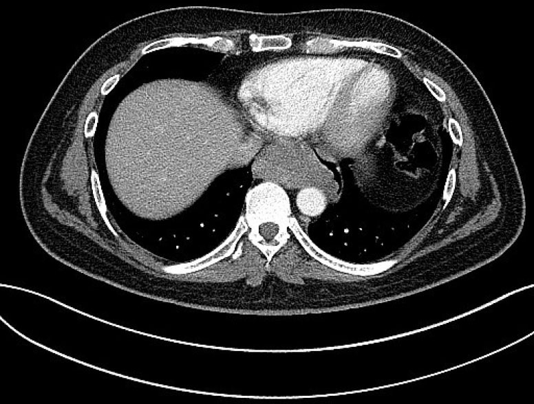
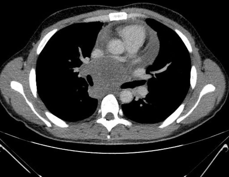
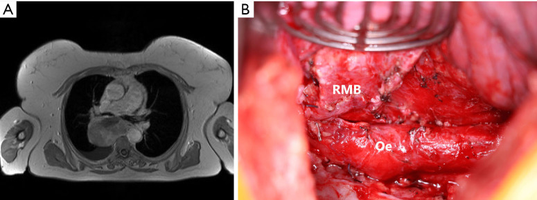
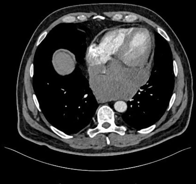
Results: Thirty-six patients (23%) had lesions located in the middle mediastinal compartment. Most common diagnoses were mediastinal cysts (n=10, 28%), metastatic lesions (n=6, 17%), lymphomas (n=5, 14%), and sarcomas (n=3, 8%). Ninetieth percentile lesion size was 73 mm. As per definition, four patients had giant middle mediastinal lesions. All these four lesions were of mesenchymal origin including oesophageal leiomyoma, synovial sarcoma, leiomyosarcoma and undifferentiated round cell sarcoma. Resection was performed through posterolateral thoracotomy or sternotomy, with or without cardiopulmonary bypass.
Conclusions: The term "giant" could be defined as a mass larger or equal to 73 mm. This definition selected specifically lesions with mesenchymal origin and may therefore guide diagnostic algorithm and patient management.
Keywords: Giant; mesenchymal tumors; middle mediastinum; sarcoma; surgery.
2023 Mediastinum. All rights reserved.
Conflict of interest statement
Conflicts of Interest: All authors have completed the ICMJE uniform disclosure form (available at https://med.amegroups.com/article/view/10.21037/med-22-49/coif). The authors have no conflicts of interest to declare.
Figures
References
-
- Fujimoto K, Hara M, Tomiyama N, et al. Proposal for a new mediastinal compartment classification of transverse plane images according to the Japanese Association for Research on the Thymus (JART) General Rules for the Study of Mediastinal Tumors. Oncol Rep 2014;31:565-72. 10.3892/or.2013.2904 - DOI - PMC - PubMed
LinkOut - more resources
Full Text Sources
Research Materials