Review
. 2023 May 8:7:27.
doi: 10.21037/med-22-53. eCollection 2023.

Multimodality imaging of mediastinal masses and mimics

Affiliations
Review

Multimodality imaging of mediastinal masses and mimics

John Matthew Archer et al. Mediastinum. .

Abstract

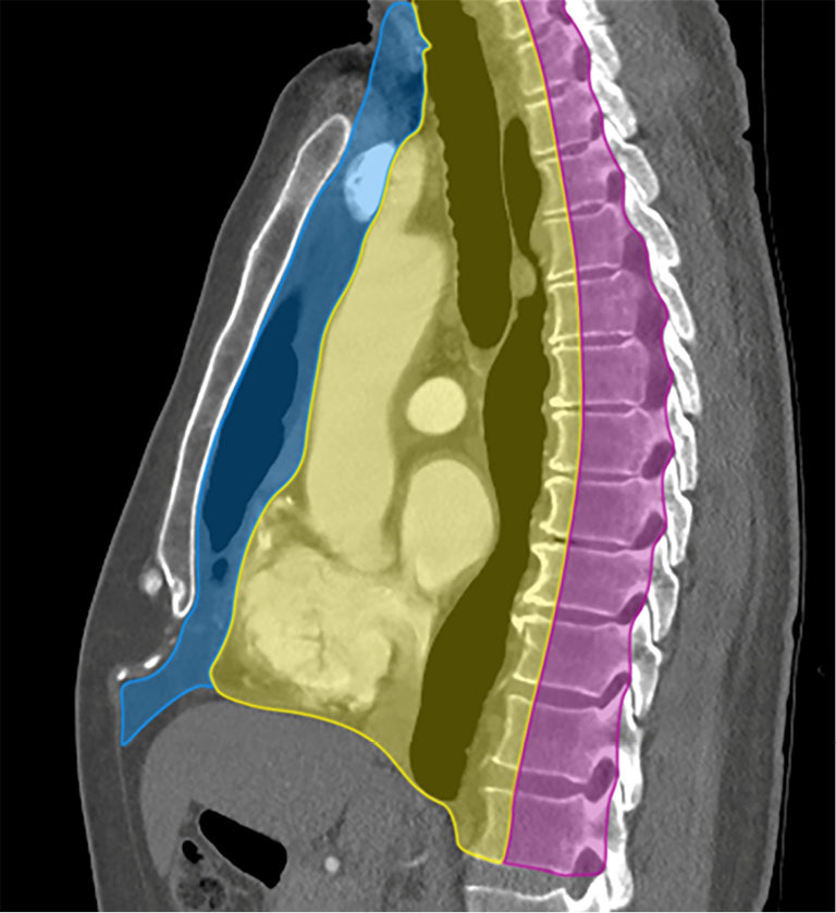
A wide variety of neoplastic and nonneoplastic conditions occur in the mediastinum. Imaging plays a central role in the evaluation of mediastinal pathologies and their mimics. Localization of a mediastinal lesion to a compartment and characterization of morphology, density/signal intensity, enhancement, and mass effect on neighboring structures can help narrow the differentials. The International Thymic Malignancy Interest Group (ITMIG) established a cross-sectional imaging-derived and anatomy-based classification system for mediastinal compartments, comprising the prevascular (anterior), visceral (middle), and paravertebral (posterior) compartments. Cross-sectional imaging is integral in the evaluation of mediastinal lesions. Computed tomography (CT) and magnetic resonance imaging (MRI) are useful to characterize mediastinal lesions detected on radiography. Advantages of CT include its widespread availability, fast acquisition time, relatively low cost, and ability to detect calcium. Advantages of MRI include the lack of radiation exposure, superior soft tissue contrast resolution to detect invasion of the mass across tissue planes, including the chest wall and diaphragm, involvement of neurovascular structures, and the potential for dynamic sequences during free-breathing or cinematic cardiac gating to assess motion of the mass relative to adjacent structures. MRI is superior to CT in the differentiation of cystic from solid lesions and in the detection of fat to differentiate thymic hyperplasia from thymic malignancy.

Keywords: Mediastinal compartments; computed tomography (CT); magnetic resonance imaging (MRI); mediastinal mass.


Conflict of interest statement

Conflicts of Interest: All authors have completed the ICMJE uniform disclosure form (available at https://med.amegroups.com/article/view/10.21037/med-22-53/coif). The authors have no conflicts of interest to declare.

Figures

Figure 1
Figure 1
ITMIG classification of mediastinal compartments bound superiorly by the thoracic inlet and inferiorly by the diaphragm. Sagittal CT with intravenous contrast shows the prevascular compartment (blue), bound anteriorly by the sternum and posteriorly by the anterior aspect of the parietal pericardium, and contains the thymus, fat, lymph nodes, and the left brachiocephalic vein. The visceral compartment (yellow) is bound anteriorly by the posterior boundaries of the prevascular compartment, and posteriorly by a vertical line 1 cm posterior to the anterior margin of each thoracic vertebral body, and contains the trachea, esophagus, lymph nodes, and vascular structures including the heart, thoracic aorta, superior vena cava, intrapericardial pulmonary arteries, and the thoracic duct. The paravertebral compartment (pink) is bound anteriorly by the posterior boundaries of the visceral compartment, and posterolaterally by a vertical line against the posterior margin of the chest wall at the lateral margin of the transverse process of the thoracic spine. ITMIG, International Thymic Malignancy Interest Group; CT, computed tomography.
Figure 2
Figure 2
Pericardial cyst. (A,B) Frontal and lateral chest radiograph shows subtle contour abnormality (arrows) with loss of the silhouette of part of the right mediastinal border termed the “silhouette sign”. (C) CT shows low attenuation consistent with pericardial cyst (arrow). CT, computed tomography.
Figure 3
Figure 3
Pericardial cyst. (A,B) MRI images show typical low signal intensity on T1-weighted (A) and high signal intensity on T2-weighted (B) images of fluid (arrow) in the pericardial cyst. MRI, magnetic resonance imaging.
Figure 4
Figure 4
Esophageal duplication cyst. (A) Contrast enhanced CT shows lesion (arrow) in the right subcarinal region with heterogeneous attenuation and could be cystic or solid. When CT is indeterminate, MRI is useful to differentiate fluid from solid mediastinal lesions. (B,C) MRI images show the visceral mediastinal lesion (arrow) with intermediate signal intensity on T1-weighted (B) and high signal intensity on T2-weighted (C) MRI images. The intermediate signal intensity on T1-weighted images can be seen with cysts with proteinaceous material. CT, computed tomography; MRI, magnetic resonance imaging.
Figure 5
Figure 5
Mature teratoma. Contrast enhanced CT shows large right mediastinal mass with heterogeneous attenuation, including calcifications (long arrow) and fat (short arrows). There is mass effect on the right atrium. CT, computed tomography.
Figure 6
Figure 6
Fat necrosis. (A) CT shows oval lesion (arrow) posterior to the gastric pull-through in a patient treated for esophageal cancer. The lesion shows predominant fat attenuation demarcated by a soft tissue rim, typical of fat necrosis. (B) FDG PET/CT shows the fat necrosis (arrow) is FDG avid due to the inflammatory component and can be misinterpreted as tumor recurrence. CT, computed tomography; FDG, fluoro-2-deoxy-D-glucose; PET, positron emission tomography.
Figure 7
Figure 7
Hemangioma. CT shows large left mediastinal mass with focus of calcification (arrow) consistent with phlebolith interspersed with soft tissue and fat. CT, computed tomography.
Figure 8
Figure 8
Carney’s triad. (A) Contrast enhanced CT shows enhancement of the left paratracheal mediastinal soft tissue mass (asterisk) due to paraganglioma and a solid lung nodule in the left upper lobe with coarse calcification (arrow) consistent with chondroma. (B) FDG PET/CT shows increased FDG uptake of the paraganglioma (asterisk). (C) Contrast-enhanced CT abdomen shows round well-circumscribed soft tissue nodule (asterisk) in the anterior wall of the stomach consistent with gastrointestinal stromal tumor. CT, computed tomography; FDG, fluoro-2-deoxy-D-glucose; PET, positron emission tomography.
Figure 9
Figure 9
Ectopic parathyroid adenoma. (A,B) Pre- and post-contrast enhanced CT show right paratracheal solid lesion (arrow) with heterogeneous enhancement. The pre-contrast CT is useful to differentiate high attenuation thyroid tissue from low attenuation parathyroid tissue. (C,D) Technetium-99m sestamibi parathyroid scintigraphy and SPECT/CT show the right parathyroid adenoma is sestamibi avid (arrow). CT, computed tomography; SPECT, single photon emission computed tomography.
Figure 10
Figure 10
Ganglioneuroma. (A,B) PA and lateral chest radiographs show elongated bilateral posterior mediastinal lesion (arrows) extending from the neck down to below the diaphragm. (C) Axial contrast enhanced CT shows bilateral paraspinal masses with low attenuation and enlargement of the right neural foramen of T4 (arrow) with extension into the spinal canal. (D) Axial T1-weighted MRI shows the mass (arrow) is heterogeneous with iso- to hyperintense signal. Extension into the spinal canal displaces and the spinal cord to the left with abnormal signal intensity within the cord due to cord compression and edema. (E) Axial T1-weighted post-contrast MRI shows the mass (arrow) enhances heterogeneously. (F) Axial T2-weighted MRI shows the mass (arrow) is heterogeneously hyperintense. (G) Fused PET/CT shows the ganglioneuroma is not FDG avid. Biopsy confirmed ganglioneuroma. PA, posterior anterior; CT, computed tomography; MRI, magnetic resonance imaging; PET, positron emission tomography; FDG, fluoro-2-deoxy-D-glucose.
Figure 11
Figure 11
Schwannoma. (A,B) PA and lateral chest radiographs show a left mediastinal mass (arrow) in the upper thorax. (C) Axial contrast enhanced CT shows the mass (arrow) is solid with heterogeneous attenuation. (D) Axial T1-weighted MRI shows the mass (arrow) is iso-intense to muscle. (E) Axial T1-weighted post-contrast MRI shows the mass (arrow) enhances heterogeneously. (F) Axial T2-weighted MRI shows the mass (arrow) is heterogeneously hyperintense with cystic areas. (G) Fused PET/CT shows the mass (arrow) is FDG avid. Biopsy confirmed schwannoma. PA, posterior anterior; CT, computed tomography; MRI, magnetic resonance imaging; PET, positron emission tomography; FDG, fluoro-2-deoxy-D-glucose.
Figure 12
Figure 12
SVC obstruction. (A) Axial contrast enhanced CT shows mediastinal adenopathy due to lung cancer narrowing the SVC (arrow) with multiple collaterals in the right anterior chest wall. (B) Coronal contrast enhanced CT shows the SVC is patent below the level of obstruction due to the adenopathy (arrow). SVC, superior vena cava; CT, computed tomography.

Similar articles

  • Diagnostic approach to the anterior/prevascular mediastinum for radiologists.
    Carter BW, Benveniste MF, Marom EM. Carter BW, et al. Mediastinum. 2019 May 27;3:18. doi: 10.21037/med.2018.12.03. eCollection 2019. Mediastinum. 2019. PMID: 35118246 Free PMC article. Review.
  • Approach to the prevascular mass.
    Tomiyama N. Tomiyama N. Mediastinum. 2019 May 8;3:17. doi: 10.21037/med.2019.04.05. eCollection 2019. Mediastinum. 2019. PMID: 35118245 Free PMC article. Review.
  • MR Imaging of Mediastinal Masses.
    Carter BW, Betancourt SL, Benveniste MF. Carter BW, et al. Top Magn Reson Imaging. 2017 Aug;26(4):153-165. doi: 10.1097/RMR.0000000000000134. Top Magn Reson Imaging. 2017. PMID: 28777164 Review.
  • [Mediastinum-new compartment classification].
    Röhrich S, Heidinger BH, Prayer F, Kifjak D, Beer L, Wassipaul C, Watzenböck M, Milos RI, Prosch H. Röhrich S, et al. Radiologie (Heidelb). 2023 Mar;63(3):154-159. doi: 10.1007/s00117-023-01115-w. Epub 2023 Jan 30. Radiologie (Heidelb). 2023. PMID: 36715717 Review. German.
  • Pediatric mediastinal masses.
    Sreedher G, Tadros SS, Janitz E. Sreedher G, et al. Pediatr Radiol. 2022 Sep;52(10):1935-1947. doi: 10.1007/s00247-022-05409-4. Epub 2022 Jun 8. Pediatr Radiol. 2022. PMID: 35674800 Review.

Cited by

References

    1. Carter BW, Tomiyama N, Bhora FY, et al. A modern definition of mediastinal compartments. J Thorac Oncol 2014;9:S97-101. 10.1097/JTO.0000000000000292 - DOI - PubMed
    1. Carter BW, Benveniste MF, Madan R, et al. ITMIG Classification of Mediastinal Compartments and Multidisciplinary Approach to Mediastinal Masses. Radiographics 2017;37:413-36. 10.1148/rg.2017160095 - DOI - PubMed
    1. Expert Panel on Thoracic Imaging ; Ackman JB, Chung JH, et al. ACR Appropriateness Criteria® Imaging of Mediastinal Masses. J Am Coll Radiol 2021;18:S37-S51. 10.1016/j.jacr.2021.01.007 - DOI - PubMed
    1. Carter BW, Okumura M, Detterbeck FC, et al. Approaching the patient with an anterior mediastinal mass: a guide for radiologists. J Thorac Oncol 2014;9:S110-8. 10.1097/JTO.0000000000000295 - DOI - PubMed
    1. Heeger AP, Ackman JB. Added Value of Magnetic Resonance Imaging for the Evaluation of Mediastinal Lesions. Radiol Clin North Am 2021;59:251-77. 10.1016/j.rcl.2020.11.001 - DOI - PubMed