Review
. 2023 Apr 12:7:25.
doi: 10.21037/med-22-50. eCollection 2023.

Spindle cell thymoma and its histological mimickers

Affiliations
Review

Spindle cell thymoma and its histological mimickers

Annikka Weissferdt. Mediastinum. .

Abstract

Spindle cell thymomas are the most common spindle cell neoplasms of the anterior mediastinum. These tumors belong to the group of thymic epithelial neoplasms and are known for their wide histomorphologic spectrum. This histological heterogeneity is the reason why unequivocal diagnosis can be challenging, especially when dealing with small biopsy material. Conversely, less conventional patterns of the tumor may also pose significant diagnostic problems in resected material and the differential diagnosis often includes other spindle cell neoplasms that are known to arise in the mediastinal cavity. These can be of variable origin and may share overlapping pathological features with spindle cell thymoma. Since spindle cell thymomas are tumors that primarily affect the adult population and predominantly arise from the thymic gland in the anterior mediastinum, this review will focus on the differential diagnosis with other spindle cell neoplasms that share similar demographic characteristics and, for the most part, originate from the anterior mediastinal compartment. These include other epithelial spindle cell tumors of thymic origin (sarcomatoid thymic carcinoma and spindle cell carcinoid tumor), mesenchymal neoplasms [solitary fibrous tumor (SFT), synovial sarcoma, and dedifferentiated liposarcoma] and various other tumors with spindle cell morphology, that may occasionally involve the anterior mediastinum. The clinical, pathological, immunohistochemical and molecular hallmarks of these lesions will be discussed and useful tips for the differential diagnosis with spindle cell thymoma will be provided.

Keywords: Mediastinum; sarcoma; spindle cell; thymic carcinoid tumor; thymic carcinoma; thymoma.


Conflict of interest statement

Conflicts of Interest: The author has completed the ICMJE uniform disclosure form (available at https://med.amegroups.com/article/view/10.21037/med-22-50/coif). AW serves as an unpaid editorial board member of Mediastinum from October 2021 to September 2023. The author has no other conflicts of interest to declare.

Figures

Figure 1
Figure 1
Spindle cell thymoma. (A) Low power view of a spindle cell thymoma showing a lobulated pattern with tumor nodules divided by thick fibrous bands (H&E, ×4); (B) the tumor cells are distributed in a sheet-like arrangement (H&E, ×10); (C) the tumor cells are spindle-shaped, have bland cytologic features and are percolated by scattered small lymphocytes (thymocytes) (H&E, ×20); (D) perivascular spaces, if present, are a good clue to the diagnosis of thymoma (H&E, ×20). H&E, hematoxylin and eosin.
Figure 2
Figure 2
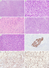
Different variants of spindle cell thymoma include (A) micronodular thymoma with lymphoid hyperplasia (H&E, ×4), (B) adenomatoid spindle cell thymoma (H&E, ×4), (C) ancient (sclerosing) thymoma (H&E, ×10), (D) angiomatoid spindle cell thymoma (H&E, ×10), (E) desmoplastic spindle cell thymoma (H&E, ×10), (F) spindle cell thymoma with papillary/pseudopapillary features (H&E, ×10), (G) spindle cell thymoma with neuroendocrine pattern (H&E, ×20), and (H) spindle cell thymoma with neural (meningothelial) pattern (H&E, ×20). H&E, hematoxylin and eosin.
Figure 3
Figure 3
Immunohistochemical phenotype of spindle cell thymoma. Tumor cells with diffuse expression of (A) pancytokeratin (×4), (B) CK5/6 (×10), (C) p40 (×4), and (D) Pax8 (polyclonal) (×10).
Figure 4
Figure 4
Spindle cell carcinoid tumor of the thymus. (A) Low power view of a thymic spindle cell carcinoid tumor characterized by tumor cells arranged in a nested pattern (H&E, ×4); (B) the tumor cells have fusiform nuclei with a salt-and-pepper chromatin pattern and low to absent mitotic activity (H&E, ×20); the tumor cells show diffuse reactivity with neuroendocrine markers, including (C) synaptophysin (×10) and (D) chromogranin A (×10). H&E, hematoxylin and eosin.
Figure 5
Figure 5
Sarcomatoid thymic carcinoma composed of (A) spindle cells arranged in a fascicular pattern (H&E, ×10); (B) individual tumor cells demonstrate a degree of cytologic atypia and conspicuous mitotic figures (H&E, ×20); by immunohistochemistry, the tumor cells are positive for (C) pancytokeratin (×20) and (D) p40 (×10).
Figure 6
Figure 6
Mediastinal SFT. (A) SFT with a characteristic “patternless pattern” (H&E, ×4); (B) keloid-like collagen is a common finding in these tumors (H&E, ×10); immunohistochemical expression of (C) CD34 (×10) and (D) STAT6 in a mediastinal SFT (×10). SFT, solitary fibrous tumor; H&E, hematoxylin and eosin.
Figure 7
Figure 7
Mediastinal synovial sarcoma. (A) Synovial sarcomas are typically highly cellular spindle cell lesions (H&E, ×4); (B) the tumor cells are monomorphic and mitotically active (H&E, ×20); (C) synovial sarcoma with dense stromal collagen (H&E, ×10); (D) biphasic synovial sarcoma characterized by glandular structures in addition to the spindle cell component (H&E, ×10); (E) poorly differentiated synovial sarcoma demonstrating small round tumor cells with rhabdoid features (H&E, ×10); (F) cytokeratin immunohistochemical expression in a biphasic synovial sarcoma is restricted to the glandular component (×10); novel markers for synovial sarcomas include (G) TLE1 (×10) and (H) SS18-SSX (×10). H&E, hematoxylin and eosin.
Figure 8
Figure 8
Mediastinal dedifferentiated liposarcoma: (A) a well differentiated liposarcoma component (right) is juxtaposed with dedifferentiated areas (left) (H&E, ×4); (B) the non-lipogenic elements may have a hemangiopericytoma-like growth pattern (H&E, ×10); cytologically, the dedifferentiated tumor may show (C) high-grade (H&E, ×10) or (D) low-grade features (H&E, ×20). H&E, hematoxylin and eosin.
Figure 9
Figure 9
Miscellaneous spindle cell neoplasms of the mediastinum. (A) Mediastinal angiosarcoma presenting as a cellular spindle cell tumor (H&E, ×10); (B) immunohistochemistry for ERG in a mediastinal angiosarcoma (×10); (C) spindle cells with a wavy configuration are characteristic for schwannomas (H&E, ×10); (D) the tumor cells of a schwannoma are diffusely positive for S100 protein (×10); (E) inflammatory myofibroblastic tumor composed of bland spindle cells admixed with an inflammatory cell infiltrate (H&E, ×10); (F) reactivity for smooth muscle antigen in a mediastinal inflammatory myofibroblastic tumor (×10). H&E, hematoxylin and eosin; ERG, erythroblast transformation specific related gene.
Figure 10
Figure 10
Miscellaneous spindle cell neoplasms of the mediastinum. (A) Sarcomatoid mesothelioma composed of pleomorphic spindle cells arranged in a haphazard pattern (H&E, ×10); (B) the tumor cells of a sarcomatoid mesothelioma are highlighted by a pancytokeratin immunostain (×10); (C) intimal sarcoma characterized by an undifferentiated cellular spindle cell proliferation with central necrosis (H&E, ×10); (D) this particular case displayed variable reactivity with smooth muscle actin by immunohistochemistry (×10). H&E, hematoxylin and eosin.

Similar articles

References

    1. Weissferdt A. Mesenchymal Tumors of the Mediastinum. In: Weissferdt A. Diagnostic Thoracic Pathology. 1st ed. Cham: Springer; 2020:972.
    1. Weissferdt A, Moran CA. The histomorphologic spectrum of spindle cell thymoma. Hum Pathol 2014;45:437-45. 10.1016/j.humpath.2012.08.025 - DOI - PubMed
    1. Ströbel P, Badve S, Chan JKC, et al. Type A thymoma. In: World Health Organization. WHO Classification of Tumours Editorial Board. Thoracic Tumours. 5th ed. Lyon: IARC Press; 2021:326-31.
    1. Nonaka D, Rosai J. Is there a spectrum of cytologic atypia in type a thymomas analogous to that seen in type B thymomas? A pilot study of 13 cases. Am J Surg Pathol 2012;36:889-94. 10.1097/PAS.0b013e31824fff50 - DOI - PubMed
    1. Vladislav IT, Gökmen-Polar Y, Kesler KA, et al. The role of histology in predicting recurrence of type A thymomas: a clinicopathologic correlation of 23 cases. Mod Pathol 2013;26:1059-64. 10.1038/modpathol.2013.49 - DOI - PubMed