Review
. 2023 Aug 27;15(8):1574-1590.
doi: 10.4240/wjgs.v15.i8.1574.

Vascular complications of chronic pancreatitis and its management

Affiliations
Review

Vascular complications of chronic pancreatitis and its management

Dinesh Walia et al. World J Gastrointest Surg. .

Abstract

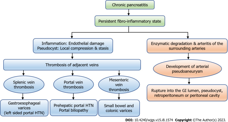
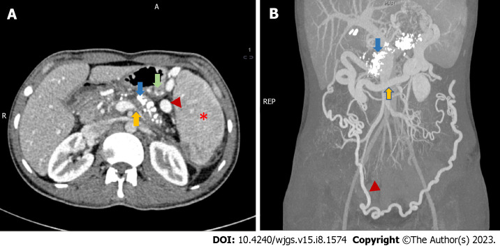
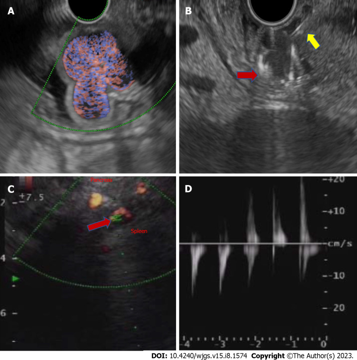
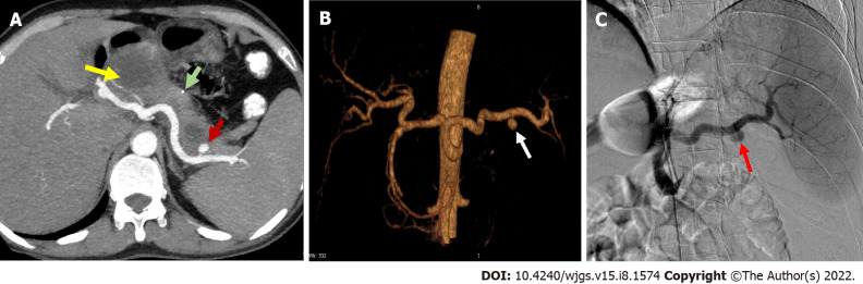
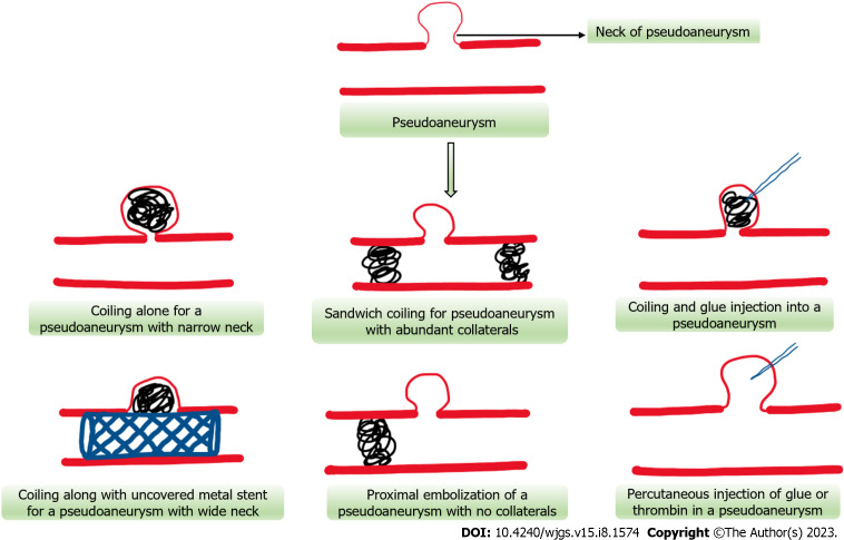
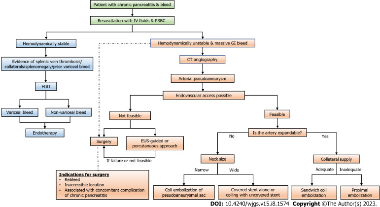
Chronic pancreatitis is a chronic fibro-inflammatory disorder of the pancreas, resulting in recurrent abdominal pain, diabetes mellitus, and malnutrition. It may lead to various other complications such as pseudocyst formation, benign biliary stricture, gastric outlet obstruction; and vascular complications like venous thrombosis, variceal and pseudoaneurysmal bleed. Development of varices is usually due to chronic venous thrombosis with collateral formation and variceal bleeding can easily be tackled by endoscopic therapy. Pseudoaneurysmal bleed can be catastrophic and requires radiological interventions including digital subtraction angiography followed by endovascular obliteration, or sometimes with a percutaneous or an endoscopic ultrasound-guided approach in technically difficult situations. Procedure-related bleed is usually venous and mostly managed conservatively. Procedure-related arterial bleed, however, may require radiological interventions.

Keywords: Chronic pancreatitis; Pseudoaneurysm; Varices; Vascular complications; Venous thrombosis.


Conflict of interest statement

Conflict-of-interest statement: All the authors report no relevant conflicts of interest for this article.

Figures

Figure 1
Figure 1
The types of vascular complications in chronic pancreatitis and their consequences. GI: Gastrointestinal; HTN: Hypertension.
Figure 2
Figure 2
Radiological features of chronic calcific pancreatitis and its complications including venous thrombosis and collaterals. A: An axial section of contrast-enhanced computed tomography (CECT) showing chronic pancreatitis with calcifications (blue arrow), attenuated splenic vein (yellow arrow), multiple perigastric collaterals (green arrow), gastrosplenic collaterals (red arrowhead) and splenomegaly (red asterisk); B: Coronal section of CECT of the same patient showing extensive pancreatic calcification (blue arrow) with dilated gastroepiploic vein (yellow arrow) and omental collaterals (red arrowhead). Courtesy: Dr Madhusudhan KS, Department of Radiodiagnosis.
Figure 3
Figure 3
Endoscopic ultrasound-guided fundal variceal obliteration, and pseudoaneurysm on endoscopic ultrasound. A: Linear endoscopic ultrasound showing the fundal varices on doppler study; B: Linear endoscopic ultrasound guided metal coil (red arrow, hyperechoic curved structure) being pushed into the varices for obliteration after the endoscopic ultrasound needle puncture (yellow arrow, hyperechoic linear structure); C: Linear endoscopic ultrasound showing an arterial pseudoaneurysm (red arrow) on doppler study; D: On power doppler mode Doppler showing an arterial waveform with bidirectional flow, classically labelled as “yin-yang” sign.
Figure 4
Figure 4
Arterial pseudoaneurysm in chronic pancreatitis. A: Axial contrast-enhanced computed tomography in a patient of chronic pancreatitis showing a pseudoaneurysm (PsA) (red arrow) arising from splenic artery along with a specks of parenchymal calcification (green arrow) and a pseudocyst (yellow arrow) in the head of pancreas; B: Reconstructed angiographic image of the same patient showing splenic artery PsA (white arrow); C: Digital subtraction angiography of the same patient showing contrast outpouching from the splenic artery suggestive of splenic artery PsA (red arrow) with contrast opacification before endovascular therapy. Courtesy: Dr Madhusudhan KS, Department of Radiodiagnosis.
Figure 5
Figure 5
Schematic representation of various endovascular and percutaneous approaches for pseudoaneurysm obliteration.
Figure 6
Figure 6
Flow diagram depicting the approach to a case of gastrointestinal bleeding in patients with chronic pancreatitis (dashed arrow suggests an alternative management strategy). CT: Computed tomography; EGD: Esophagogastroduodenoscopy; GI: Gastrointestinal; PRBC: Packed red blood cell; EUS: Endoscopic ultrasound.

References

    1. Vege SS, Chari ST. Chronic Pancreatitis. N Engl J Med. 2022;386:869–878. - PubMed
    1. Olesen SS, Krauss T, Demir IE, Wilder-Smith OH, Ceyhan GO, Pasricha PJ, Drewes AM. Towards a neurobiological understanding of pain in chronic pancreatitis: mechanisms and implications for treatment. Pain Rep. 2017;2:e625. - PMC - PubMed
    1. Murruste M, Kirsimägi Ü, Kase K, Veršinina T, Talving P, Lepner U. Complications of chronic pancreatitis prior to and following surgical treatment: A proposal for classification. World J Clin Cases. 2022;10:7808–7824. - PMC - PubMed
    1. Agarwal S, Sharma S, Gunjan D, Singh N, Kaushal K, Poudel S, Anand A, Gopi S, Mohta S, Sonika U, Saraya A. Natural course of chronic pancreatitis and predictors of its progression. Pancreatology. 2020;20:347–355. - PubMed
    1. Ramsey ML, Conwell DL, Hart PA. Complications of Chronic Pancreatitis. Dig Dis Sci. 2017;62:1745–1750. - PMC - PubMed