

Preferential FGF18/FGFR activity in pseudoglandular versus canalicular stage human lung fibroblasts
- PMID: 37701781
- PMCID: PMC10493313
- DOI: 10.3389/fcell.2023.1220002
Preferential FGF18/FGFR activity in pseudoglandular versus canalicular stage human lung fibroblasts
Abstract
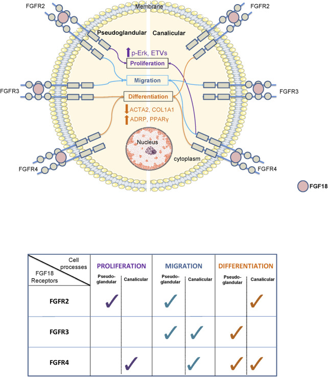
Fibroblast growth factor (FGF) signaling is necessary for proper lung branching morphogenesis, alveolarization, and vascular development. Dysregulation of FGF activity has been implicated in various lung diseases. Recently, we showed that FGF18 promotes human lung branching morphogenesis by regulating mesenchymal progenitor cells. However, the underlying mechanisms remain unclear. Thus, we aimed to determine the role of FGF18 and its receptors (FGFR) in regulating mesenchymal cell proliferation, migration, and differentiation from pseudoglandular to canalicular stage. We performed siRNA assays to identify the specific FGFR(s) associated with FGF18-induced biological processes. We found that FGF18 increased proliferation and migration in human fetal lung fibroblasts (HFLF) from both stages. FGFR2/FGFR4 played a significant role in pseudoglandular stage. HFLF proliferation, while FGFR3/FGFR4 were involved in canalicular stage. FGF18 enhanced HFLF migration through FGFR2 and FGFR4 in pseudoglandular and canalicular stage, respectively. Finally, we provide evidence that FGF18 treatment leads to reduced expression of myofibroblast markers (ACTA2 and COL1A1) and increased expression of lipofibroblast markers (ADRP and PPARγ) in both stages HFLF. However, the specific FGF18/FGFR complex involved in this process varies depending on the stage. Our findings suggest that in context of human lung development, FGF18 tends to associate with distinct FGFRs to initiate specific biological processes on mesenchymal cells.
Keywords: FGF18; FGFR; lung development; mesenchyme; progenitor cells.
Copyright © 2023 Belgacemi, Cherry, El Alam, Frauenpreis, Glass, Bellusci, Danopoulos and Al Alam.
Conflict of interest statement
The authors declare that the research was conducted in the absence of any commercial or financial relationships that could be construed as a potential conflict of interest.
Figures


Similar articles
-
FGF9 and FGF18 in idiopathic pulmonary fibrosis promote survival and migration and inhibit myofibroblast differentiation of human lung fibroblasts in vitro.Am J Physiol Lung Cell Mol Physiol. 2016 Apr 1;310(7):L615-29. doi: 10.1152/ajplung.00185.2015. Epub 2016 Jan 15. Am J Physiol Lung Cell Mol Physiol. 2016. PMID: 26773067
-
FGF18 promotes human lung branching morphogenesis through regulating mesenchymal progenitor cells.Am J Physiol Lung Cell Mol Physiol. 2023 Apr 1;324(4):L433-L444. doi: 10.1152/ajplung.00316.2022. Epub 2023 Feb 15. Am J Physiol Lung Cell Mol Physiol. 2023. PMID: 36791060 Free PMC article.
-
Up-regulation of the fibroblast growth factor 8 subfamily in human hepatocellular carcinoma for cell survival and neoangiogenesis.Hepatology. 2011 Mar;53(3):854-64. doi: 10.1002/hep.24099. Epub 2011 Feb 11. Hepatology. 2011. PMID: 21319186
-
FGFR inhibitors: Effects on cancer cells, tumor microenvironment and whole-body homeostasis (Review).Int J Mol Med. 2016 Jul;38(1):3-15. doi: 10.3892/ijmm.2016.2620. Epub 2016 May 31. Int J Mol Med. 2016. PMID: 27245147 Free PMC article. Review.
-
Divergent fibroblast growth factor signaling pathways in lung fibroblast subsets: where do we go from here?Am J Physiol Lung Cell Mol Physiol. 2015 Oct 15;309(8):L751-5. doi: 10.1152/ajplung.00298.2015. Epub 2015 Sep 4. Am J Physiol Lung Cell Mol Physiol. 2015. PMID: 26342090 Review.
Cited by
-
Mechanisms and Therapeutic Potential of Myofibroblast Transformation in Pulmonary Fibrosis.J Respir Biol Transl Med. 2025 Mar;2(1):10001. doi: 10.70322/jrbtm.2025.10001. Epub 2025 Mar 7. J Respir Biol Transl Med. 2025. PMID: 40190620 Free PMC article.
-
Morphological and molecular aspects of lung development.Histol Histopathol. 2025 Apr;40(4):411-430. doi: 10.14670/HH-18-807. Epub 2024 Sep 4. Histol Histopathol. 2025. PMID: 39344418 Review.
References
Grants and funding
LinkOut - more resources
Full Text Sources
Research Materials
Miscellaneous