. 2023 Sep 1;16(9):dmm050090.
doi: 10.1242/dmm.050090. Epub 2023 Sep 13.

Stromal matrix directs corneal fibroblasts to re-express keratocan after injury and transplantation

Affiliations

Stromal matrix directs corneal fibroblasts to re-express keratocan after injury and transplantation

Ana C Acosta et al. Dis Model Mech. .

Abstract

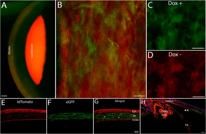
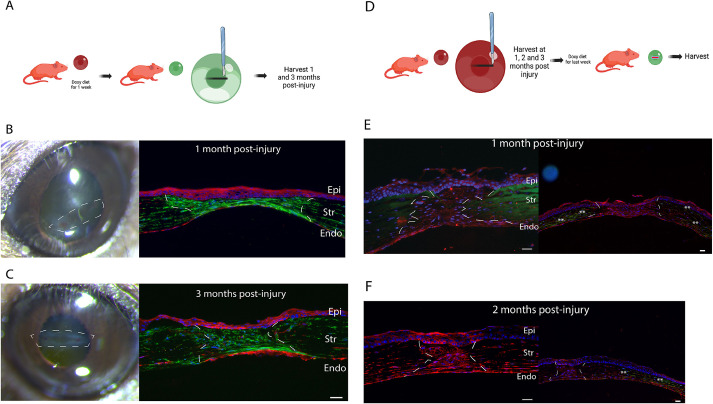
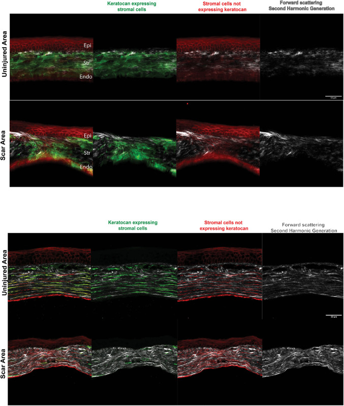
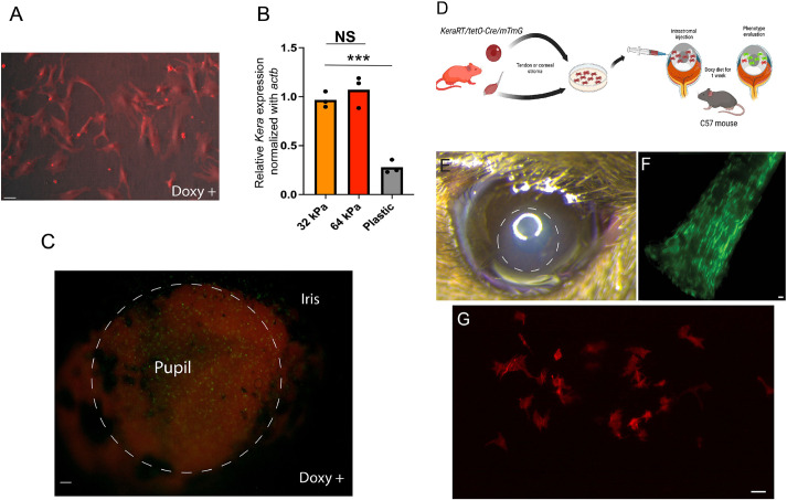
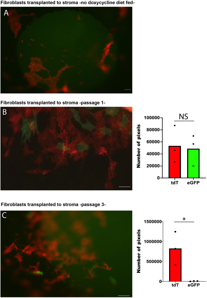
Every tissue has an extracellular matrix (ECM) with certain properties unique to it - the tissue 'niche' - that are necessary for normal function. A distinct specific population of quiescent keratocan-expressing keratocytes populate the corneal stroma during homeostasis to maintain corneal function. However, during wound healing, when there is alteration of the niche conditions, keratocytes undergo apoptosis, and activated corneal fibroblasts and myofibroblasts attempt to restore tissue integrity and function. It is unknown what the fate of activated and temporary fibroblasts and myofibroblasts is after the wound healing process has resolved. In this study, we used several strategies to elucidate the cellular dynamics of corneal wound healing and the fate of corneal fibroblasts. We injured the cornea of a novel mouse model that allows cell-lineage tracing, and we transplanted a cell suspension of in vitro-expanded corneal fibroblasts that could be tracked after being relocated into normal stroma. These transplanted fibroblasts regained expression of keratocan in vivo when relocated to a normal stromal niche. These findings suggest that transformed fibroblasts maintain plasticity and can be induced to a keratocyte phenotype once relocated to an ECM with normal signaling ECM.

Keywords: Keratocan; Keratocyte; Stroma; Transplantation.


Conflict of interest statement

Competing interests E.M.E. is a consultant with GSK. The remaining authors declare no competing or financial interests.

Figures

Fig. 1.
Fig. 1.
A triple transgenic conditional KeraRT/tetO-Cre/mTmG mouse strain. (A) The corneal stroma shows the presence of keratocan-expressing keratocytes (green) in the stroma of mouse eye after 1 week of oral doxycycline diet. (B) eGFP expression is limited to keratocan-expressing keratocytes only; epithelial cells do not express eGFP (green) and maintain their tdTomato (red) expression, as noted in this flat-mount image. (C,D) eGFP expression was noted in animals fed doxycycline (C) and was absent in a control group not fed a doxycycline-supplemented diet (D). (E-G) In this histology section, a few cells in the stroma did not express keratocan (marked by asterisks). The majority of cells expressed keratocan (F,G). Epi, epithelium; Str, stroma; Endo, endothelium. (H) Expression of eGFP by keratocytes started at the peripheral cornea (shown by asterisks) and was absent in the limbus. Images are representative of seven different eyes. Scale bars: 50 µm (A,E-H); 25 µm (B-D).
Fig. 2.
Fig. 2.
The keratocyte lineage is involved in tissue repair post injury but stromal cells in maturing scars (up to 2 months after injury) are not keratocytes. (A) The first strategy was to trace keratocyte lineage after the penetrating stromal injury by first labeling keratocytes with eGFP (doxycycline diet for 1 week) and then creating an injury. (B,C) Cells in the injury were derived from the keratocyte lineage as shown by eGFP expression in repaired/regenerated stroma at 1 month (B) and 3 months (C) post injury. (D) The second strategy was to evaluate the phenotype of cells present within maturing scars after the penetrating stromal injury by feeding mice a doxycycline diet for the last week before harvesting. At 1 and 2 months after the penetrating injury, cells did not express eGFP, suggesting that they were not keratocytes. (E) At 1 month post injury (n=6), cells present in the repaired stroma did not express keratocan. However, keratocytes were noted in the peripheral cornea outside the repaired injury area (eGFP expression shown by asterisks). (F) At 2 months after injury (n=6), cells present in the scar still did not express keratocan. White lines indicate scars. Images are representative of at least three experimental repeats from 16 mice. Epi, epithelium; Str, stroma; Endo, endothelium. Scale bars: 25 µm (B,C, right; E,F).
Fig. 3.
Fig. 3.
Imaging of mature corneal scars using SHG and fluorescence microscopy in a freshly enucleated adult I-KeramTmG eye mounted for imaging shows keratocytes in the repairing stroma 3 months after injury. A doxycycline diet was given 1 week prior to injury to assess eGFP expression in scars. Top: uninjured corneas had a predominance of keratocytes embedded within the corneal stroma, with few cells expressing tdTomato. Second-harmonic generation (SHG) imaging (gray, right) showed horizontally well-aligned and organized collagen fibrils/lamellae in the stroma. The scar area showed a mixture of keratocytes and tdTomato-expressing cells in the scar tissue, which was disorganized in an oblique and vertical array. Similarly, collagen fibrils/lamellae were disorganized (n=4). Bottom: tissue sections showed keratocytes in the repairing stroma 3 months after injury. Uninjured corneas had a predominance of keratocytes embedded within the corneal stroma (green and red signals), with a minimal number of cells expressing tdTomato (red). Cells were parallel to the basement and Descemet's membranes. SHG imaging (gray, right) showed well-aligned and horizontally organized collagen stromal fibrils/lamellae. Scars had a mixture of keratocytes and tdTomato-expressing cells oriented in a disorganized fashion. Similarly, collagen fibrils/lamellae were disorganized (n=6). Epi, epithelium; Str, stroma; Endo, endothelium. Scale bars: 50 µm.
Fig. 4.
Fig. 4.
Keratocan expression is lost during standard in vitro expansion in plastic culture dishes with serum stimulation but maintained or reacquired if cells are in their unique stromal niche. (A) Freshly passed fibroblasts expanded in vitro under serum stimulation shut down keratocan expression, despite maintaining a dendritic morphology (n=3). (B) Stromal cells downregulated keratocan expression when cultured in a stiffer matrix; plastic dishes in this case (n=3). In contrast, keratocytes expressed eGFP ex vivo – in serum medium supplemented with doxycycline – when maintained within the stromal matrix. NS, not significant; ***P=0.0003 (two-tailed unpaired t-test). (C) The image shows induced cells (eGFP expression) that are more noticeable in the region of the pupil 24 h after exposure to doxycycline in the culture medium (n=5). (D) Expanded tenocytes – known to express keratocan in vivo – or corneal-derived fibroblasts can be expanded in vitro and transplanted to the stromal matrix to evaluate whether keratocan is expressed. (E) An area of corneal edema (dashed circle) developed immediately after cell injection; the edema resolved in a few days and corneal transparency returned to normal (n=7). (F) eGFP expression in tendons in vivo after 1 week of oral doxycycline diet (n=6). (G) However, expanded tenocytes failed to express keratocan (eGFP) when transplanted to the stromal matrix (n=5). Scale bars: 25 µm (A,C,F,G).
Fig. 5.
Fig. 5.
Fibroblasts revert to a keratocyte phenotype after transplantation. (A) Transplanted cells in mice not fed with a doxycycline-supplemented diet did not express eGFP. (B) High-magnification microscopy shows the presence of keratocytes (eGFP-expressing cells) 1 week after transplantation into a normal stromal niche in mice fed with a doxycycline-supplemented diet (passage 1 cells) (n=9). (C) Very few cells expressed keratocan once corneal fibroblasts were expanded for a longer time in vitro (passage 3) (n=3). The graphs (right) show quantification of the number of image pixels for tdTomato (tdT)- and eGFP-expressing cells. NS, not significant; *P<0.05 (two-tailed paired t-test). Scale bars: 50 µm.

References

    1. Beales, M. P., Funderburgh, J. L., Jester, J. V. and Hassell, J. R. (1999). Proteoglycan synthesis by bovine keratocytes and corneal fibroblasts: maintenance of the keratocyte phenotype in culture. Invest. Ophthalmol. Vis. Sci. 40, 1658-1663. - PubMed
    1. Birk, D. E. and Trelstad, R. L. (1984). Extracellular compartments in matrix morphogenesis: collagen fibril, bundle, and lamellar formation by corneal fibroblasts. J. Cell Biol. 99, 2024-2033. 10.1083/jcb.99.6.2024 - DOI - PMC - PubMed
    1. Call, M., Elzarka, M., Kunesh, M., Hura, N., Birk, D. E. and Kao, W. W. (2019). Therapeutic efficacy of mesenchymal stem cells for the treatment of congenital and acquired corneal opacity. Mol. Vis. 25, 415-426. - PMC - PubMed
    1. Cogswell, D., Sun, M., Greenberg, E., Margo, C. E. and Espana, E. M. (2021). Creation and grading of experimental corneal scars in mice models. Ocul. Surf. 19, 53-62. 10.1016/j.jtos.2020.11.008 - DOI - PMC - PubMed
    1. Espana, E. M. and Birk, D. E. (2020). Composition, structure and function of the corneal stroma. Exp. Eye Res. 198, 108137. 10.1016/j.exer.2020.108137 - DOI - PMC - PubMed

Publication types

Substances