Review
. 2023 Sep 13;10(1):11.
doi: 10.1186/s40348-023-00167-1.

Autoimmune lymphoproliferative immunodeficiencies (ALPID) in childhood: breakdown of immune homeostasis and immune dysregulation

Affiliations
Review

Autoimmune lymphoproliferative immunodeficiencies (ALPID) in childhood: breakdown of immune homeostasis and immune dysregulation

Vasil Toskov et al. Mol Cell Pediatr. .

Abstract

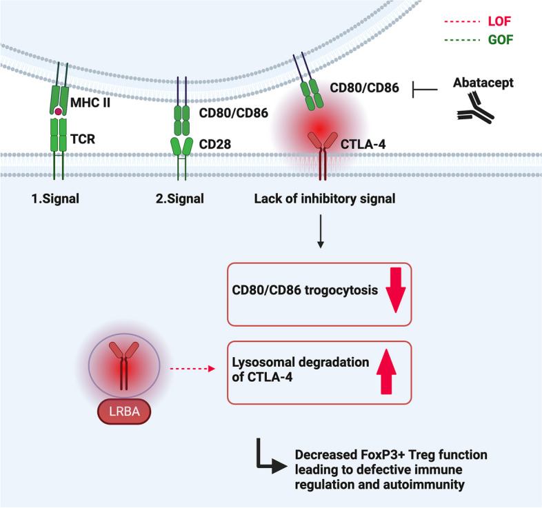
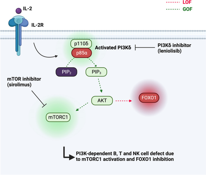
Many inborn errors of immunity (IEI) manifest with hallmarks of both immunodeficiency and immune dysregulation due to uncontrolled immune responses and impaired immune homeostasis. A subgroup of these disorders frequently presents with autoimmunity and lymphoproliferation (ALPID phenotype). After the initial description of the genetic basis of autoimmune lymphoproliferative syndrome (ALPS) more than 20 years ago, progress in genetics has helped to identify many more genetic conditions underlying this ALPID phenotype. Among these, the majority is caused by a group of autosomal-dominant conditions including CTLA-4 haploinsufficiency, STAT3 gain-of-function disease, activated PI3 kinase syndrome, and NF-κB1 haploinsufficiency. Even within a defined genetic condition, ALPID patients may present with staggering clinical heterogeneity, which makes diagnosis and management a challenge. In this review, we discuss the pathophysiology, clinical presentation, approaches to diagnosis, and conventional as well as targeted therapy of the most common ALPID conditions.

Keywords: Autoimmune lymphoproliferative immunodeficiencies; Immune dysregulation; Inborn errors of immunity; Pathogenesis; Targeted therapy.


Conflict of interest statement

The authors declare no competing interests.

Figures

Fig. 1
Fig. 1
Illustration of the Fas-FasL pathway. Fas (CD95) is a trimeric receptor of the tumor TNF-R family and after binding of the Fas ligand (FasL, CD95L), recruits the adaptor FADD, which in turn forms the so-called DISC together with pro-caspase-8 and pro-caspase-10. Following activation, caspase-8 and caspase-10 then initiate the extrinsic apoptotic pathway leading to proteolysis, DNA degradation, and apoptosis. Mutations in the genes responsible for the Fas-FasL signaling cascade (FAS, FASLG, FADD, CASP10) lead to the development of ALPS. Hallmarks of the disease are increased ALPS biomarkers (Vitamin B12 and sFasL), as well as a massive expansion of double-negative T cells (DNT). Increased AKT/mTOR activation leads to DNT hyperproliferation and can be inhibited via mTOR inhibitors such as sirolimus (rapamycin)
Fig. 2
Fig. 2
The two-step process of T cell activation. To counteract it, CTLA-4 in endosomes reaches the cell surface, outcompetes CD28, and binds and downregulates CD80/CD86 in a process called trogocytosis. LRBA acts intracellularly to stabilize and protect intracellular CTLA-4 from lysosomal degradation, thus maintaining the pool of available CTLA-4. Both in CTLA-4 haploinsufficiency and LRBA deficiency, lack of CTLA-4 (either due to decreased translation or increased lysosomal degradation) limits the function of regulatory T cells. Abatacept is a soluble CTLA-4 immunoglobulin fusion protein (Fc-region of human IgG1 linked to the extracellular domain of CTLA-4), which mimics CTLA-4 function and can be used successfully as a targeted therapy in both conditions
Fig. 3
Fig. 3
Illustration of the JAK/STAT pathway. After binding to their receptor, cytokines such as IL-6 activate an associated Janus Kinase (JAK), which upon phosphorylation of its tyrosine residues recruits and phosphorylates the STAT3 transcription regulator. Phosphorylated STAT3 in turn forms homo- or heterodimers which translocate into the nucleus and impact the transcription of cytokine-responsive genes. In STAT3 GOF, the signaling pathway can lead to increased phosphorylation, altered dimer formation, as well as changes in gene expression. Targeting molecules which are part of the STAT3 pathway leads to improved STAT3 GOF disease control, e.g., disruption of the IL-6/IL-6R interaction via the anti-IL-6R monoclonal antibody tocilizumab. Another strategy is the inhibition of JAK by jakinibs such as ruxolitinib
Fig. 4
Fig. 4
Illustration of the NF-κB pathway. The NF-κB1 transcriptional factor (p50 and its precursor p105) is active upon dimerization (p50:p65) in the cytosol. At rest, NF-κB dimers are bound to inhibitory IκB proteins. After activation (here via PRR signaling), IκB proteins are phosphorylated by the IκB kinase (IKK) complex, which releases the NF-κB dimers. p65-containing heterodimers can then translocate into the nucleus and regulate gene expression. Heterozygous loss-of-function mutations in NFKB1 are associated with reduced protein levels of the p105 and/or p50 subunit and lead to the development of a complex immunodeficiency
Fig. 5
Fig. 5
Illustration of the PI3Kδ pathway. PI3Kδ is activated through a variety of receptors (shown here is activation via IL-2 and its associated receptor). PI3Kδ typically consists of a catalytic subunit (p110δ) and a regulatory subunit (p85α) and leads to the generation of phosphatidylinositol 3,4,5-trisphosphate (PIP3) by phosphorylating its precursor phosphatidylinositol 4,5-bisphosphate (PIP2), both located in the cell membrane. Downstream PIP3 signaling is mediated by intracellular enzymes, such as the serine/threonine kinase AKT, which phosphorylates the FOXO transcription factors inactivating them, as well as regulators of the mTOR complex 1 (mTORC1), which is in turn activated. Increased activity of the PI3Kδ pathway leads to APDS. Patients with APDS are responsive to mTOR inhibition. More targeted approaches include selective PI3Kδ inhibitors, such as leniolisib or idealisib

References

    1. Farmand S, Baumann U, von Bernuth H, et al. Interdisziplinäre AWMF-Leitlinie zur Diagnostik von primären Immundefekten (S2k) Klin Pädiatrie. 2011;223:378–385. doi: 10.1055/s-0031-1287835. - DOI - PubMed
    1. Walter JE, Ayala IA, Milojevic D. Autoimmunity as a continuum in primary immunodeficiency. Curr Opin Pediatr. 2019;31:851–862. doi: 10.1097/mop.0000000000000833. - DOI - PMC - PubMed
    1. Rieux-Laucat F, Deist FL, Hivroz C, et al. Mutations in Fas associated with human lymphoproliferative syndrome and autoimmunity. Science. 1995;268:1347–1349. doi: 10.1126/science.7539157. - DOI - PubMed
    1. Fisher GH, Rosenberg FJ, Straus SE, et al. Dominant interfering fas gene mutations impair apoptosis in a human autoimmune lymphoproliferative syndrome. Cell. 1995;81:935–946. doi: 10.1016/0092-8674(95)90013-6. - DOI - PubMed
    1. Kuehn HS, Ouyang W, Lo B, et al. Immune dysregulation in human subjects with heterozygous germline mutations in CTLA4. Science. 2014;345:1623–1627. doi: 10.1126/science.1255904. - DOI - PMC - PubMed

LinkOut - more resources