. 2024 Feb 1;147(2):649-664.
doi: 10.1093/brain/awad313.

Trazodone rescues dysregulated synaptic and mitochondrial nascent proteomes in prion neurodegeneration

Affiliations

Trazodone rescues dysregulated synaptic and mitochondrial nascent proteomes in prion neurodegeneration

Hector Albert-Gasco et al. Brain. .

Abstract

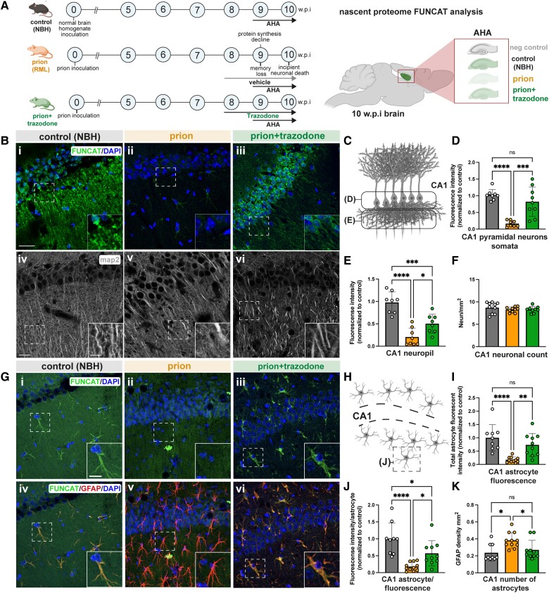
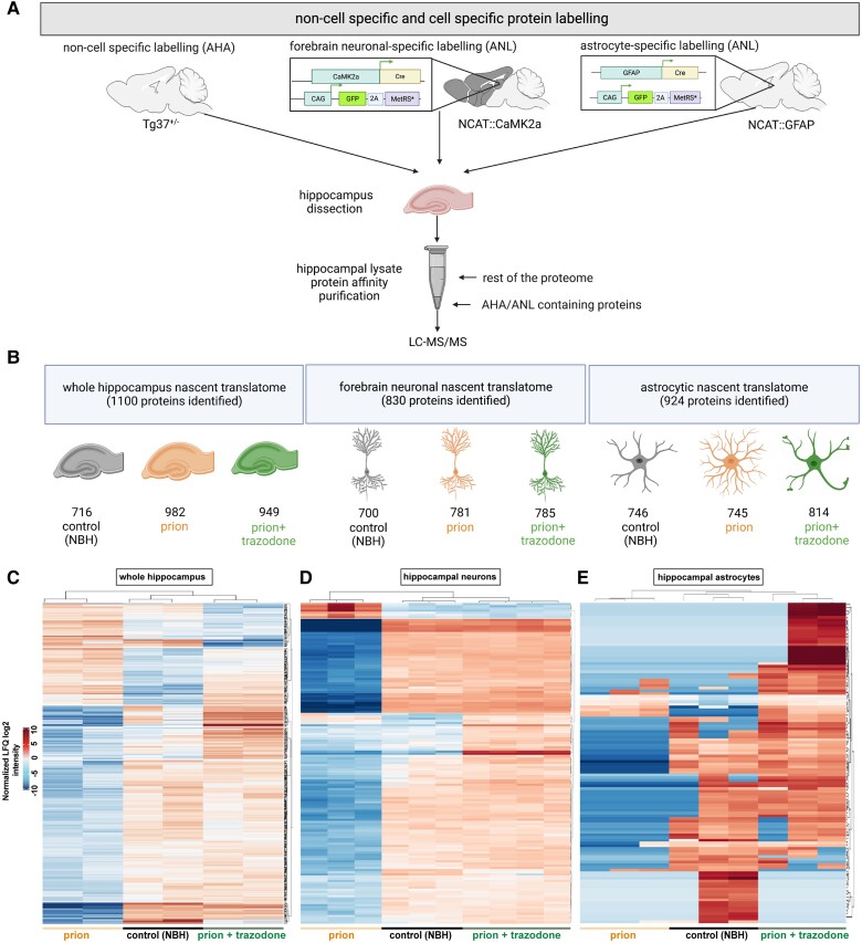
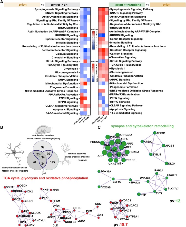
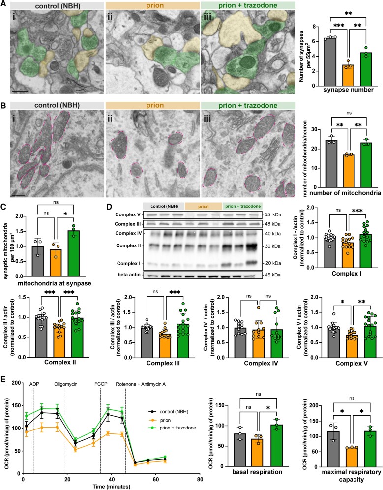
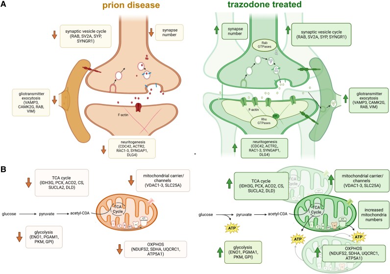
The unfolded protein response (UPR) is rapidly gaining momentum as a therapeutic target for protein misfolding neurodegenerative diseases, in which its overactivation results in sustained translational repression leading to synapse loss and neurodegeneration. In mouse models of these disorders, from Alzheimer's to prion disease, modulation of the pathway-including by the licensed drug, trazodone-restores global protein synthesis rates with profound neuroprotective effects. However, the precise nature of the translational impairment, in particular the specific proteins affected in disease, and their response to therapeutic UPR modulation are poorly understood. We used non-canonical amino acid tagging (NCAT) to measure de novo protein synthesis in the brains of prion-diseased mice with and without trazodone treatment, in both whole hippocampus and cell-specifically. During disease the predominant nascent proteome changes occur in synaptic, cytoskeletal and mitochondrial proteins in both hippocampal neurons and astrocytes. Remarkably, trazodone treatment for just 2 weeks largely restored the whole disease nascent proteome in the hippocampus to that of healthy, uninfected mice, predominantly with recovery of proteins involved in synaptic and mitochondrial function. In parallel, trazodone treatment restored the disease-associated decline in synapses and mitochondria and their function to wild-type levels. In conclusion, this study increases our understanding of how translational repression contributes to neurodegeneration through synaptic and mitochondrial toxicity via depletion of key proteins essential for their function. Further, it provides new insights into the neuroprotective mechanisms of trazodone through reversal of this toxicity, relevant for the treatment of neurodegenerative diseases via translational modulation.

Keywords: UPR/ISR; mitochondria; nascent proteome; neurodegeneration; synapses; translational repression; trazodone.


Conflict of interest statement

The authors report no competing interests.

Figures

Figure 1
Figure 1
Trazodone treatment restores repressed protein synthesis rates in prion-diseased hippocampal CA1 neurons and astrocytes. (A) Schematic showing prion disease progression and timing of L-azidohomoalanine (AHA) labelling and trazodone treatment in tg37+/− mice (left). Schematic of sagittal section of the mouse brain (right) showing hippocampal region analysed by fluorescent non-canonical amino acid tagging (FUNCAT). (B) Representative maximal projection images of the (iiii) nascent neuronal proteome labelled by FUNCAT/DAPI from summated planes within stack (40 × 0.34 μm); and (ivvi) MAP2-stained dendrites in CA1 for all conditions, showing marked reduction during prion disease and restoration by trazodone. (C) Schematic of the two regions of CA1 pyramidal neurons analysed. Fluorescence intensity quantification of newly synthesized proteins in (D) CA1 pyramidal neuron somata and (E) neuropil for all conditions calculated from summated stacks. (F) Neuronal counts in CA1 determined by NeuN staining and quantification. (G) Representative images of the (iiii) nascent proteome in astrocytes labelled by FUNCAT/DAPI from single deep sections within 40 μm slice and (ivvi) merged FUNCAT/GFAP/DAPI signal in control, prion and prion+trazodone, showing marked reduction during prion disease and restoration by trazodone. (H) Schematic showing location of CA1 astrocytes analysed. (I) Total fluorescence intensity for total CA1 astrocytes and (J) fluorescence intensity/astrocyte calculated from summated stacks for all conditions. (K) Numbers of astrocytes in CA1 region per condition. Scale bars in B(i) = 25 μm; B(iv) = 20 μm; B(iv, inset) = 5 μm and G(i, inset) = 10 μm. *P < 0.05, **P < 0.001, ***P < 0.0001, ****P < 0.00001. NBH = normal brain homogenate; ns = non-significant; w.p.i. = weeks post-inoculation.
Figure 2
Figure 2
Trazodone restores the nascent proteomes of neurons, astrocytes and whole hippocampus in prion-diseased brains. (A) Schematic showing experimental steps for bioorthogonal non-canonical amino acid tagging (BONCAT) analysis: and L-azidohomoalanine (AHA)/L-azidonorleucine (ANL) labelling of the mouse brain, hippocampal dissection, followed by affinity purification of AHA/ANL-labelled proteins. (B) Total numbers of proteins detected in control [normal brain homogenate (NBH)], prion and prion + trazodone treatment groups by liquid chromatography-tandem mass spectrometry in the whole hippocampus, in neurons and in astrocytes, respectively. (C) Hierarchical heat map by log2 normalized label-free quantification (LFQ) intensity from whole hippocampus, (D) neurons and (E) astrocytes, showing clustering of proteins and mice per experimental group. Differentially expressed significant proteins, P < 0.05.
Figure 3
Figure 3
Trazodone treatment restores specific subsets of proteins in the whole hippocampus and cell-specifically, in neurons and astrocytes. (A) Volcano plots of whole hippocampus nascent proteomes comparing (i) prion with control [normal brain homogenate (NBH)] nascent proteomes; (ii) prion + trazodone with prion; and (iii) control (NBH) with prion + trazodone. (B) Volcano plots of hippocampal forebrain neuronal nascent proteomes comparing (i) prion with control (NBH) nascent proteomes; (ii) prion + trazodone with prion; and (iii) control (NBH) with prion + trazodone. (C) Volcano plots of hippocampal astrocytic nascent proteomes comparing prion with (i) control (NBH) nascent proteomes; (ii) prion + trazodone with prion; and (iii) control (NBH) with prion + trazodone. Differentially expressed significant proteins, P < 0.05.
Figure 4
Figure 4
Synapse remodelling and mitochondrial function are the predominantly dysregulated pathways in prion disease and are restored by trazodone treatment. (A) Heat maps of significant pathways showing differences in z-score of prion (orange) versus control [normal brain homogenate (NBH)] (grey) nascent proteomes (left), and heat maps of significant pathways showing differences in z-score of prion + trazodone (green) versus prion (orange) nascent proteomes (right). (B) Venn diagram of overlapping prion + trazodone nascent proteomes from whole hippocampus, neurons and astrocytes, compared to prion alone (blue line highlights overlap). (C) Significant clusters of overlapping prion + trazodone nascent proteomes, determined by metascape-string analysis, of significantly upregulated proteins compared to prion alone. The proteins clustered to synapse and cytoskeleton remodelling, and to the TCA cycle, glycolysis and oxidative phosphorylation. P < 0.05 for significant proteins. pv = −log10P-value.
Figure 5
Figure 5
Trazodone treatment restores synapse and mitochondrial numbers and function in prion disease. (A) Representative scanning electron microscopy (SEM) images from (i) control [normal brain homogenate (NBH)]; (ii) prion; and (iii) prion + trazodone mice from CA1 stratum radiatum. Dendrites are pseudo-coloured in yellow and axons in green. Synapse number quantification of the hippocampal CA1 neuropil shows a decrease in prion disease (orange bars) and partial but significant restoration with trazodone treatment (green bars). (B) Representative SEM images from (i) control (NBH); (ii) prion; and (iii) prion + trazodone from CA1 pyramidal neuron somata, with mitochondria perimeter coloured in pink. The numbers of mitochondria/neuron are reduced in prion disease (orange bars) and restored to wild-type levels with trazodone treatment (green bars). (C) Mitochondria counted within synapses increase with trazodone treatment. (D) Immunoblots and quantification (bar graph, right) of oxidative phosphorylation proteins (I, II, III, IV and V) showing restoration with trazodone treatment. (E) Mitochondrial stress test on isolated mitochondria from control (NBH), prion and prion + trazodone treatment groups at 10 weeks post-inoculation (wpi) as before (Fig. 1A), showing oxygen consumption rates (OCRs) from 0 to 65 min, with time points of stressor addition (ADP, oligomycin, FCCP and rotenone + antimycin A, respectively) labelled. Basal respiration was defined as the (initial) OCR value minus (rotenone + antimycin A) OCR value; maximal respiratory capacity was defined as the OCR value after FCCP injection minus (rotenone + antimycin A) OCR. *P < 0.05, **P < 0.001, ***P < 0.0001. Scale bars in A(i) and B(i) = 1 μm.
Figure 6
Figure 6
Trazodone treatment in prion disease. Schematic summarizing pathway and major proteins downregulated in the nascent proteome during prion disease and recovery after trazodone treatment for (A) synapses and (B) mitochondria.

Similar articles

Cited by

References

    1. Hoozemans JJM, Veerhuis R, Van Haastert ES, et al. . The unfolded protein response is activated in Alzheimer’s disease. Acta Neuropathol. 2005;110(2):165–172. - PubMed
    1. Hoozemans JJM, Van Haastert ES, Eikelenboom JJM, de Vos RAI, Rozemuller JM, Scheper W. Activation of the unfolded protein response in Parkinson’s disease. Biochem Biophys Res Commun. 2007;354:707–711. - PubMed
    1. Hoozemans JJM, Van Haastert ES, Nijholt DAT, Rozemuller AJM, Eikelenboom P, Scheper W. The unfolded protein response is activated in pretangle neurons in Alzheimer ‘s disease hippocampus. Am J Pathol. 2009;174:1241–1251. - PMC - PubMed
    1. Nijholt DAT, Van Haastert ES, Rozemuller AJM, Scheper W, Hoozemans JJM. The unfolded protein response is associated with early tau pathology in the hippocampus of tauopathies. J Pathol. 2012;226:693–702. - PubMed
    1. Stutzbach LD, Xie SX, Naj AC, et al. . The unfolded protein response is activated in disease-affected brain regions in progressive supranuclear palsy and Alzheimer’s disease. Acta Neuropathol Commun. 2013;1:31. - PMC - PubMed

Publication types