. 2023 Sep 15;9(37):eadi2687.
doi: 10.1126/sciadv.adi2687. Epub 2023 Sep 13.

PARP14 is a PARP with both ADP-ribosyl transferase and hydrolase activities

Affiliations

PARP14 is a PARP with both ADP-ribosyl transferase and hydrolase activities

Nina Đukić et al. Sci Adv. .

Abstract

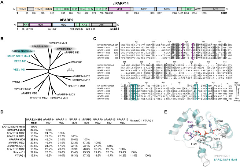
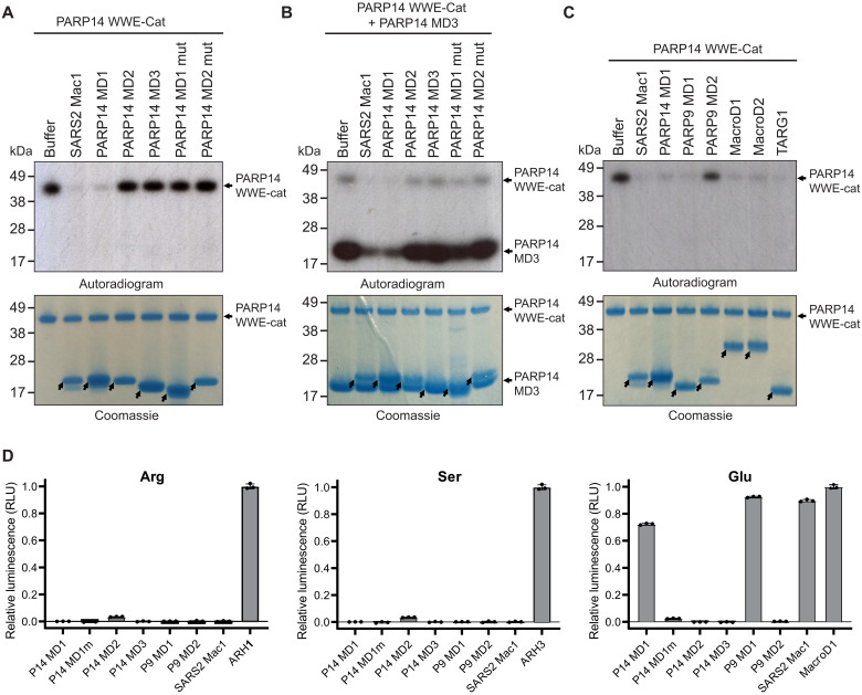
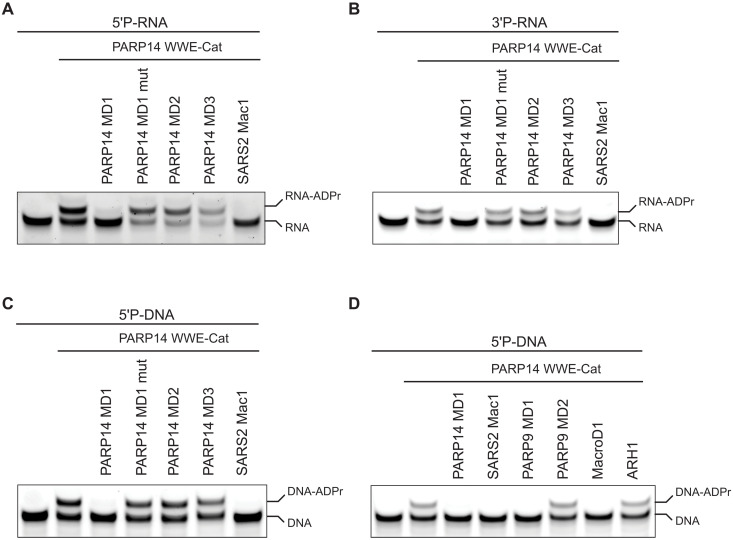
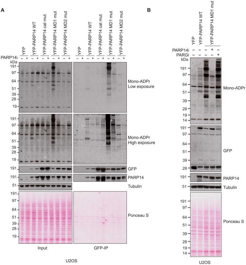
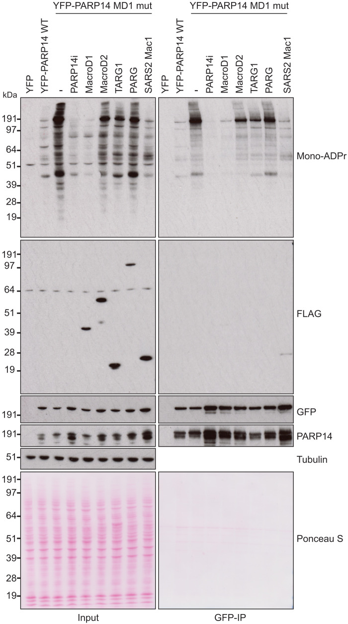
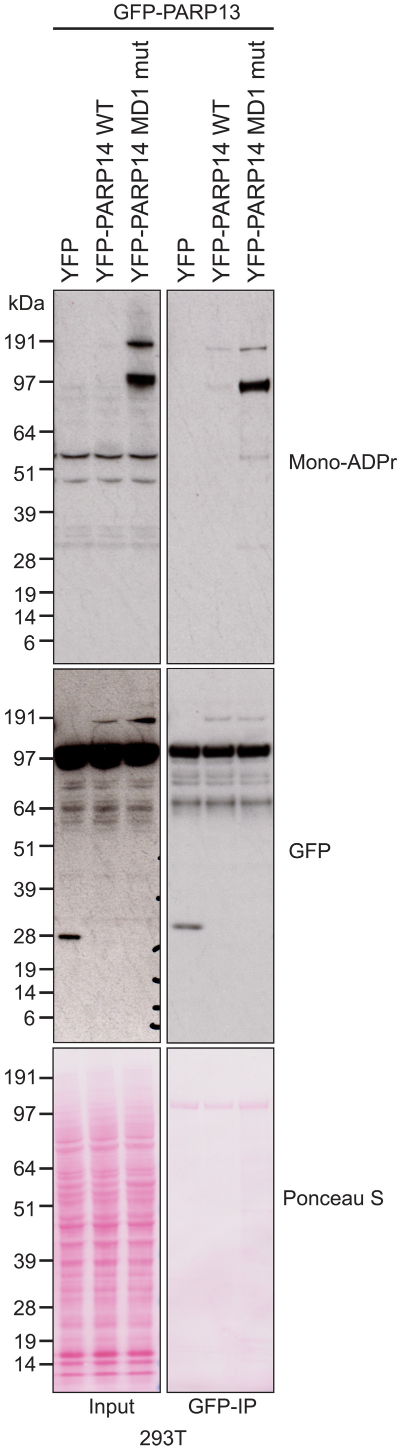
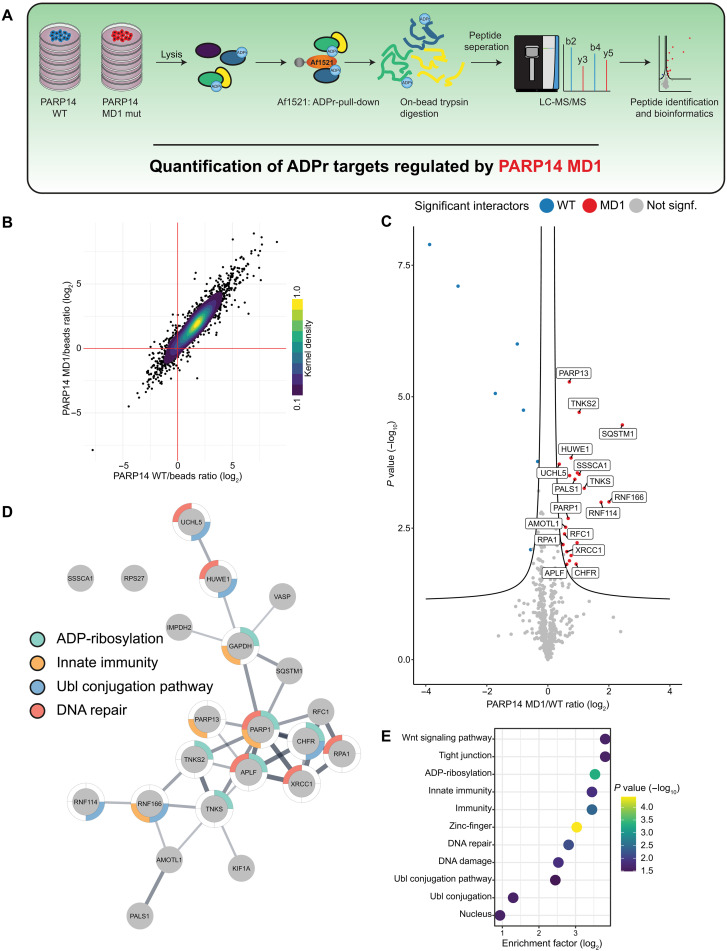
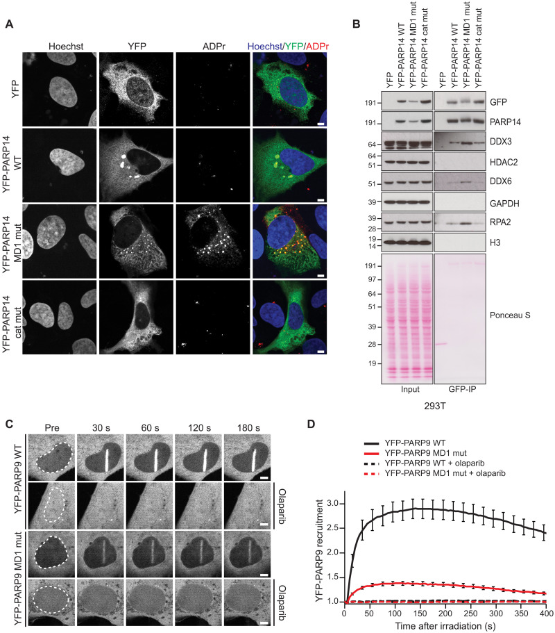
PARP14 is a mono-ADP-ribosyl transferase involved in the control of immunity, transcription, and DNA replication stress management. However, little is known about the ADP-ribosylation activity of PARP14, including its substrate specificity or how PARP14-dependent ADP-ribosylation is reversed. We show that PARP14 is a dual-function enzyme with both ADP-ribosyl transferase and hydrolase activity acting on both protein and nucleic acid substrates. In particular, we show that the PARP14 macrodomain 1 is an active ADP-ribosyl hydrolase. We also demonstrate hydrolytic activity for the first macrodomain of PARP9. We reveal that expression of a PARP14 mutant with the inactivated macrodomain 1 results in a marked increase in mono(ADP-ribosyl)ation of proteins in human cells, including PARP14 itself and antiviral PARP13, and displays specific cellular phenotypes. Moreover, we demonstrate that the closely related hydrolytically active macrodomain of SARS2 Nsp3, Mac1, efficiently reverses PARP14 ADP-ribosylation in vitro and in cells, supporting the evolution of viral macrodomains to counteract PARP14-mediated antiviral response.


Figures

Fig. 1.
Fig. 1.. PARP14 and PARP9 macrodomain 1 are similar to SARS2 Mac1.
(A) Domain architecture of human PARP14 and PARP9. (B) Unrooted phylogenetic tree of human and mouse macrodomains including of PARP9, 14, and 15 and viral macrodomains (highlighted in cyan). (C) Multiple sequence alignment showing conservation of catalytic residues (magenta-framed) and residues involved in ADP-ribose coordination (cyan-framed) of human PARP14 and PARP9 macrodomains in comparison to SARS2 Nsp3 Mac1. Numbers on top of the residues refer to human PARP14 MD1. (D) Pairwise sequence identity comparison of SARS2 Nsp3 Mac1 and human macrodomains. (E) Crystal structure overlay of PARP14 MD1 [Protein Data Bank (PDB) ID: 3Q6Z] and SARS2 Nsp3 Mac1 (PDB ID: 7KQP) both in complex with ADP-ribose (root mean square deviation of 1.02 Å over 214 Cα). The catalytic residues are highlighted in magenta.
Fig. 2.
Fig. 2.. PARP14 and PARP9 MD1 reverse glutamate-linked PARP14 auto- and trans-ADPr.
(A) PARP14 WWE-CAT was auto–ADP-ribosylated using NAD+ spiked with 32P NAD+. The ADP-ribosyl hydrolysis activity of PARP14 MD1, MD2, MD3, MD1mut (G823E), MD2mut (G1044E), and SARS2 Nsp3 Mac1 (SARS2 Mac1) was assessed upon incubation with automodified PARP14 WWE-CAT. (B) PARP14 WWE-CAT and PARP14 MD3 were auto– and trans–ADP-ribosylated, respectively, using NAD+ spiked with 32P NAD+. The trans-ADPr hydrolysis activity of PARP14 MD1, MD2, MD3, MD1mut, MD2mut, and SARS2 Mac1 was assessed upon incubation with the trans-modified PARP14 MD3 and auto-modified PARP14 WWE-CAT. (C) PARP14 WWE-CAT was auto–ADP-ribosylated using NAD+ spiked with 32P NAD+. The ADP-ribosyl hydrolysis activity of SARS2 Mac1, PARP14 MD1, PARP9 MD1, PARP9 MD2, MacroD1, MacroD2, and TARG1 was determined upon incubation with automodified PARP14 WWE-CAT. Samples in (A to C) were analyzed by Coomassie brilliant blue staining and autoradiography. The arrows show the position of the indicated macrodomains. (D) Hydrolysis of arginine-, serine-, and glutamate-linked mono(ADP-ribosyl)ation on synthetic peptides by PARP14 MD1, PARP14 MD1mut, PARP14 MD2, PARP14 MD3, PARP9 MD1, PARP9 MD2, and SARS2 Mac1. Briefly, the released ADP-ribose was converted by NUDT5 to adenosine 5′-monophosphate (AMP), which subsequently was detected by luminescence using the AMP-Glo assay (Promega). Samples are background-corrected and normalized to the positive control, ARH1 for arginine, ARH3 for serine, and MacroD1 for glutamate. The data represent means ± SD measured in triplicates.
Fig. 3.
Fig. 3.. PARP14 and PARP9 MD1 reverse ADPr of ssRNA and ssDNA.
(A) ssRNA with 5′ phosphate and 3′ cyanine 3 (Cy3), (B) ssRNA with 3′ phosphate and 5′ Cy3, and (C) ssDNA with 5′ phosphate and 3′ Cy3 were ADP-ribosylated using PARP14 WWE-CAT. Subsequently, the ADPr was hydrolyzed by treating the modified oligos with PARP14 MD1, MD1mut, MD2, MD3, or SARS2 Mac1. (D) ssDNA with 5′ phosphate and 3′ Cy3 was ADP-ribosylated using PARP14 WWE-CAT. Following, the ADP ribose modification was hydrolyzed by subjecting the ADP-ribosylated oligo to PARP14 MD1, SARS2 Mac1, PARP9 MD1 and MD2, MacroD1, or ARH1.
Fig. 4.
Fig. 4.. PARP14 ADPr is reversed by its own macrodomain 1.
(A) U2OS cells were transfected with the indicated plasmids in the presence or absence of PARP14 inhibitor (PARP14i). Cell lysates and green fluorescent protein (GFP)–immunoprecipitations (GFP-IPs) were examined by Western blotting using the indicated antibodies. (B) U2OS cells were transfected with the indicated plasmids in the presence of PARP14 or PARG inhibitor. Cell lysates were examined by Western blotting with the indicated antibodies. For all blots, tubulin was used as a loading control.
Fig. 5.
Fig. 5.. MacroD1 and SARS2 Mac1 can reverse PARP14 ADPr.
U2OS cells were transfected with the indicated PARP14 plasmid together in the presence or absence of PARP14i or with FLAG-tagged MacroD1, MacroD2, TARG1, PARG, or SARS2 Mac1. Cell lysates and GFP-IPs were examined by Western blotting using the indicated antibodies. Tubulin was used as a loading control.
Fig. 6.
Fig. 6.. PARP14 acts to add and remove ADPr on PARP13.
293T cells were cotransfected with GFP-PARP13 and the indicated YFP plasmid. Cell lysates and GFP-IPs were examined by Western blotting using the indicated antibodies. Ponceau S staining was used to indicate equal loading.
Fig. 7.
Fig. 7.. MS identification of ADP-ribosylated proteins regulated by PARP14 macrodomain 1.
(A) Overview of the experimental design. (B) Scatter plot analysis of proteins enriched specifically by the ADPr-binding Af1521 macrodomain. The mean difference in abundance between proteins enriched by the Af1521 compared to control beads for cells expressing WT PARP14 is plotted against cells expressing PARP14 MD1 mutant. The color scale represents the normalized kernel density estimation of the data. (C) Analysis of proteins specifically enriched for ADPr in cells expressing PARP14 MD1 mutant compared to WT. The volcano plot shows the sample conditions (x axis), plotted against the corresponding P value resulting from two-tailed Student’s t testing (y axis). Proteins significantly down- or up-regulated [false discovery rate (FDR) < 0.05, s0 = 0.1] are represented as blue or red dots, respectively. N = 5. (D) STRING network visualizing functional interactions (edges) between proteins (nodes) significantly enriched in cells expressing PARP14 MD1 mutant over WT. The thickness of the edges corresponds to their score, and the default STRING clustering confidence score cutoff of 0.4 was used to determine whether two nodes were functionally related. Proteins were colored according to the UniProt keyword displayed on the figure legend if it was significantly enriched. (E) Gene set enrichment analysis showing UniProt keywords functionally enriched in the MD1-specific network [highlighted in (D)]. Significantly enriched terms were determined by Fisher exact testing, testing whether categorical terms found in the MD1-specific network were functionally enriched over terms found in the background, which was defined as all proteins significantly enriched by Af1521 over bead controls. Terms were determined to be significant with a Benjamini-Hochberg multiple-hypotheses corrected P value <0.05. The terms are ranked by their functional enrichment over the background in descending order and colored by their corresponding Benjamini-Hochberg–adjusted P values.
Fig. 8.
Fig. 8.. PARP14 and PARP9 macrodomain 1 regulates various cellular functions.
(A) Confocal images of U2OS cells expressing YFP or YFP-PARP14 WT, MD1 mutant (MD1 mut), or catalytically inactive mutant (cat mut) stained with Hoechst (blue) and YFP (green) and for ADPr (poly/mono) (red). Scale bars, 5 μm. (B) 293T cells were cotransfected with the indicated YFP plasmid. Cell lysates and GFP-IPs were examined by Western blotting using the indicated antibodies. Ponceau S staining was used to indicate equal loading. (C) Confocal images and (D) recruitment kinetics of YFP-PARP9 and YFP-PARP9 macrodomain 1 mutant (MD1 mut) to sites of laser irradiation in the absence or presence of 1 μM olaparib. Scale bars, 5 μm.

References

    1. R. Gupte, Z. Liu, W. L. Kraus, PARPs and ADP-ribosylation: Recent advances linking molecular functions to biological outcomes. Genes Dev. 31, 101–126 (2017). - PMC - PubMed
    1. J. Groslambert, E. Prokhorova, I. Ahel, ADP-ribosylation of DNA and RNA. DNA Repair (Amst) 105, 103144 (2021). - PMC - PubMed
    1. S. Vyas, I. Matic, L. Uchima, J. Rood, R. Zaja, R. T. Hay, I. Ahel, P. Chang, Family-wide analysis of poly(ADP-ribose) polymerase activity. Nat. Commun. 5, 4426 (2014). - PMC - PubMed
    1. B. Lüscher, I. Ahel, M. Altmeyer, A. Ashworth, P. Bai, P. Chang, M. Cohen, D. Corda, F. Dantzer, M. D. Daugherty, T. M. Dawson, V. L. Dawson, S. Deindl, A. R. Fehr, K. L. H. Feijs, D. V. Filippov, J.-P. Gagné, G. Grimaldi, S. Guettler, N. C. Hoch, M. O. Hottiger, P. Korn, W. L. Kraus, A. Ladurner, L. Lehtiö, A. K. L. Leung, C. J. Lord, A. Mangerich, I. Matic, J. Matthews, G.-L. Moldovan, J. Moss, G. Natoli, M. L. Nielsen, M. Niepel, F. Nolte, J. Pascal, B. M. Paschal, K. Pawłowski, G. G. Poirier, S. Smith, G. Timinszky, Z.-Q. Wang, J. Yélamos, X. Yu, R. Zaja, M. Ziegler, ADP-ribosyltransferases, an update on function and nomenclature. FEBS J. 289, 7399–7410 (2022). - PMC - PubMed
    1. O. Leidecker, J. J. Bonfiglio, T. Colby, Q. Zhang, I. Atanassov, R. Zaja, L. Palazzo, A. Stockum, I. Ahel, I. Matic, Serine is a new target residue for endogenous ADP-ribosylation on histones. Nat. Chem. Biol. 12, 998–1000 (2016). - PMC - PubMed