. 2023 Nov:67:102866.
doi: 10.1016/j.redox.2023.102866. Epub 2023 Sep 4.

Hemin and iron increase synthesis and trigger export of xanthine oxidoreductase from hepatocytes to the circulation

Affiliations

Hemin and iron increase synthesis and trigger export of xanthine oxidoreductase from hepatocytes to the circulation

Evan R DeVallance et al. Redox Biol. 2023 Nov.

Abstract

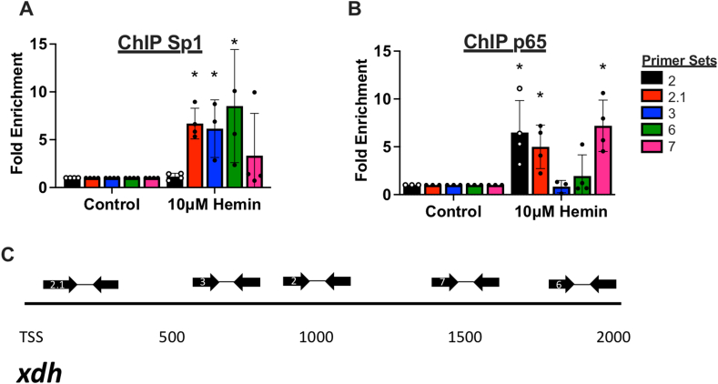
We recently reported a previously unknown salutary role for xanthine oxidoreductase (XOR) in intravascular heme overload whereby hepatocellular export of XOR to the circulation was identified as a seminal step in affording protection. However, the cellular signaling and export mechanisms underpinning this process were not identified. Here, we present novel data showing hepatocytes upregulate XOR expression/protein abundance and actively release it to the extracellular compartment following exposure to hemopexin-bound hemin, hemin or free iron. For example, murine (AML-12 cells) hepatocytes treated with hemin (10 μM) exported XOR to the medium in the absence of cell death or loss of membrane integrity (2.0 ± 1.0 vs 16 ± 9 μU/mL p < 0.0001). The path of exocytosis was found to be noncanonical as pretreatment of the hepatocytes with Vaculin-1, a lysosomal trafficking inhibitor, and not Brefeldin A inhibited XOR release and promoted intracellular XOR accumulation (84 ± 17 vs 24 ± 8 hemin vs 5 ± 3 control μU/mg). Interestingly, free iron (Fe2+ and Fe3+) induced similar upregulation and release of XOR compared to hemin. Conversely, concomitant treatment with hemin and the classic transition metal chelator DTPA (20 μM) or uric acid completely blocked XOR release (p < 0.01). Our previously published time course showed XOR release from hepatocytes likely required transcriptional upregulation. As such, we determined that both Sp1 and NF-kB were acutely activated by hemin treatment (∼2-fold > controls for both, p < 0.05) and that silencing either or TLR4 with siRNA prevented hemin-induced XOR upregulation (p < 0.01). Finally, to confirm direct action of these transcription factors on the Xdh gene, chromatin immunoprecipitation was performed indicating that hemin significantly enriched (∼5-fold) both Sp1 and NF-kB near the transcription start site. In summary, our study identified a previously unknown pathway by which XOR is upregulated via SP1/NF-kB and subsequently exported to the extracellular environment. This is, to our knowledge, the very first study to demonstrate mechanistically that XOR can be specifically targeted for export as the seminal step in a compensatory response to heme/Fe overload.

Keywords: Heme; Hemin; Hepatocytes; Iron; Oxidants; Xanthine oxidoreductase.


Conflict of interest statement

Declaration of competing interest The authors declare that they have no known competing financial interests or personal relationships that could have appeared to influence the work reported in this paper.

Figures

Fig. 1
Fig. 1
Hemin-induced release of XOR from hepatocytes. A) Plasma XO activity in wild-type C57BL6J mice following vehicle or hemin injection measured by HPLC. B) AML12 cells treated with increasing concentration of hemin for 24 h. Media was collected to measure XO activity by HPLC on the left Y-axis (black) while the corresponding cells had cell death measured by NucGreen fluorescent probe normalized to 0.5 mM triton-X positive control on the right Y-axis (Red). C) AML 12 cells treated with 10 μM hemin for 24 h. Media was collected and XO activity measured. D) AML12 cells were treated with 10 μM hemin±the necroptosis inhibitor necrostatin 1 for 24 h. Media was collected and XO activity measured. E) AML 12 cells were treated with 10 μM hemin or 0.17 mM Triton-X for 24 h. Media was collected and XO activity measured. F) Primary hepatocytes were isolated from wild-type mice and plated in cell culture plates then treated with 10 μM hemin for 24 h. Media was collected and XO activity measured. Individual data points shown with mean ± SD depicted; significance indicated as * p < 0.05, **p < 0.01, ***p < 0.001, ****p < 0.0001. XO, xanthine oxidase; HPLC, high performance liquid chromatography. (For interpretation of the references to colour in this figure legend, the reader is referred to the Web version of this article.)
Fig. 2
Fig. 2
Hemin-induced upregulation of XOR in hepatocytes. A) XOR expression measured by Western blot in AML 12 cells 24-h treatment with 10 μM hemin. B) XDH mRNA measured by qPCR in AML12 cells following 6-h treatment with 10 μM hemin. Individual data points shown with mean ± SD depicted; significance indicated as * p < 0.05, **p < 0.01. XDH, xanthine dehydrogenase; G3PDH, glyceraldehyde 3-phosphate dehydrogenase; XOR, xanthine oxidoreductase.
Fig. 3
Fig. 3
Effect of trafficking inhibitors on XOR release from hepatocytes. A) AML 12 cells treated with 10 μM hemin±Brefeldin A or Vacuolin-1. Media was collected and XO activity measured by HPLC. B) AML 12 cells treated with 10 μM hemin±Brefeldin A or Vacuolin-1. Cells were then collected and XO activity measured by HPLC. Individual data points shown with mean ± SD depicted; significance indicated as **p < 0.01, ****p < 0.0001. XO, xanthine oxidase; HPLC, high performance liquid chromatography.
Fig. 4
Fig. 4
Iron sulfate-induced XOR release and XDH gene transcription. A) AML 12 cells treated with 10 μM FeSO4 +/− 10 μM PPIX for 24 h. Media was collected and XO activity measured by HPLC. B) XDH mRNA measured by qPCR in AML12 cells treated for 6 h with 10 μM FeSO4 +/− 10 μM PPIX. Individual data points shown with mean ± SD depicted; significance indicated as ***p < 0.001, ****p < 0.0001. XO, xanthine oxidase; FeSO4, Ferrous sulfate; PPIX, protoporphyrin IX; XDH, xanthine dehydrogenase; G3PDH, glyceraldehyde 3-phosphate dehydrogenase; HPLC, high performance liquid chromatography.
Fig. 5
Fig. 5
Iron chelators DTPA and Uric acid prevent hemin and iron sulfate-induced XOR release. A&B) AML 12 cells treated with 10 μM hemin or 10 μM FeSO4 for 24 h ± 20 μM DTPA or 20 μM UA. Media was collected and XO activity measured by HPLC. C) AML 12 cells treated with 10 μM hemin ± 10 μM HPX for 24 h. Media collected and XO activity measured. D) EPR assessment of ascorbate radical production for 10 μM hemin±treatment with DTPA and UA with representative spectra (i-blank, ii-10μM hemin, iii-10μM hemin+ 20 μM DTPA, iv-10μM hemin+ 20 μM UA). Individual data points shown with mean ± SD depicted; significance indicated as ***p < 0.001, ****p < 0.0001. XO, xanthine oxidase; FeSO4, Ferrous sulfate; DTPA, diethylenetriamine pentaacetate; UA, uric acid; HPX, hemopexin; EPR, electron paramagnetic resonance; HPLC, high performance liquid chromatography.
Fig. 6
Fig. 6
Transcriptional regulation of XOR by p65 and Sp1. A) AML 12 cells treated 10 μM hemin and H2O2 measured by CBA. B&C) In-cell and traditional Western blot detection of phosphorylated p65 in AML 12 cells treated with 10 μM hemin for 2 h. D&E) In-cell and traditional Western blot detection of phosphorylated Sp1 in AML 12 cells treated with 10 μM hemin for 2 h. F) AML12 cells transfected with scrambled or targeted siRNA for 48 h and then treated with 10 μM hemin. In-cell western was used to detect XOR expression. G) XDH mRNA measured by qPCR in AML12 cells transfected with scrambled and targeted siRNAs for 48 h followed by 6-h treatment with 10 μM hemin. H) AML12 cells transfected with scrambled or targeted siRNA for 48 h and then treated with 10 μM hemin for 24 h. Media was collected and XO activity measured by HPLC. Individual data points shown with mean ± SD depicted, significance indicated as * p < 0.05, **p < 0.01, ***p < 0.001, ****p < 0.0001. XDH, xanthine dehydrogenase; XOR, xanthine oxidoreductase; XO, xanthine oxidase; siScr, scrambled non-targeting siRNA.
Fig. 7
Fig. 7
Chromatin Immunoprecipitation of XDH. AML12 cells treated with hemin for 6 h were processed by ActiveMotif high-sensitivity ChIP-IT kit and DNA pulled down via A) Sp1 or B) p65 primary antibody. Immunoprecipitated DNA was analyzed by qPCR with C) 5 primer sets spanning the XDH promoter region. Individual data points shown with mean ± SD depicted; significance indicated as * p < 0.05.
Supplemental Figure 1
Vesicle Transport inhibitors prevent hemin-induced XO release. AML 12 cells treated with 10μM hemin +/- Nocodazole or Endosidin 2. Media was collected and XO activity measured by HPLC. Individual data points shown with mean ± SD depicted; significance indicated as * p<0.05. XO, xanthine oxidase; HPLC, high performance liquid chromatography.
Supplemental Fig. 2
Supplemental Fig. 2
Ferric chloride induces XO release. AML 12 cells treated with 10 μM FeCl +/− PPIX for 24 h. Media was collected and XO activity measure by HPLC. Individual data points shown with mean ± SD depicted; significance indicated as **p < 0.01. XO, xanthine oxidase; HPLC, high performance liquid chromatography; FeCl, ferric chloride; PPIX, protoporphyrin IX.
Supplemental Fig. 3
Supplemental Fig. 3
Hemin effects on transcription factor activation at 24 h and siRNA efficiency. A-C) AML 12 cells treated with 10 μM hemin for 24 h with H2O2 production measure by CBA, p65 phosphorylation and Sp1 phosphorylation measured by western blot. D&E) AML12 cells transfected with siRNA against p65 or Sp1 for 48 h then collected and knockdown efficiency measured by western blot.
Supplemental Fig. 4
Supplemental Fig. 4
Whole blots for representative images. A) Full blots for representative images used in Fig. 6C&E. B) Replicates of the representative in-cell western used in Fig. 6B&D. C) Replicates of representative in-cell western used in Fig. 6F.

References

    1. Battelli M.G., Bolognesi A., Polito L. Pathophysiology of circulating xanthine oxidoreductase: new emerging roles for a multi-tasking enzyme. Biochim. Biophys. Acta. 2014;1842(9):1502–1517. - PubMed
    1. Kelley E.E. Dispelling dogma and misconceptions regarding the most pharmacologically targetable source of reactive species in inflammatory disease, xanthine oxidoreductase. Arch. Toxicol. 2015;89(8):1193–1207. - PubMed
    1. B R. Ueber dis oxidative und dis vermeindlich synthetische Bildung von Haurnsaure in Rinderleberauszug. Zeitsohr f Physiol Chem. 1905;43:497.
    1. S W. Die Ueberführung von Nucleïnbasen in Harnsäure durch die sauerstoffübertragende Wirkung von Gewebsauszügen. Arch Ges Physiol. 1899;(76):192–203.
    1. Fz S. Uber das verhalten der kulmilch gegen methylenblau und seine verwendung zur unterscheidung von ungekochter und gekochter milch. Utersuch Nahrungs- u Genussmittel. 1902;(5):1113–1121.

Publication types

LinkOut - more resources