. 2023 Aug 1;37(15-16):760-777.
doi: 10.1101/gad.350597.123. Epub 2023 Sep 13.

Uncovering a mammalian neural-specific poly(A) binding protein with unique properties

Affiliations

Uncovering a mammalian neural-specific poly(A) binding protein with unique properties

Sahil Sharma et al. Genes Dev. .

Abstract

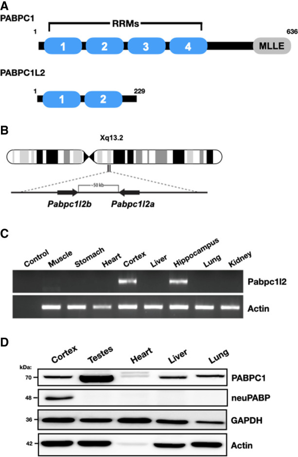
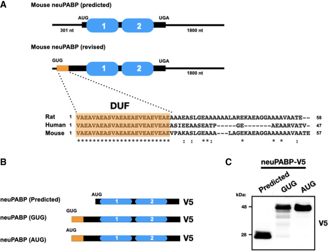
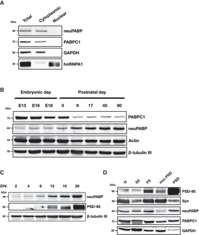
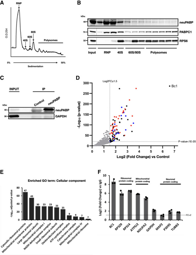
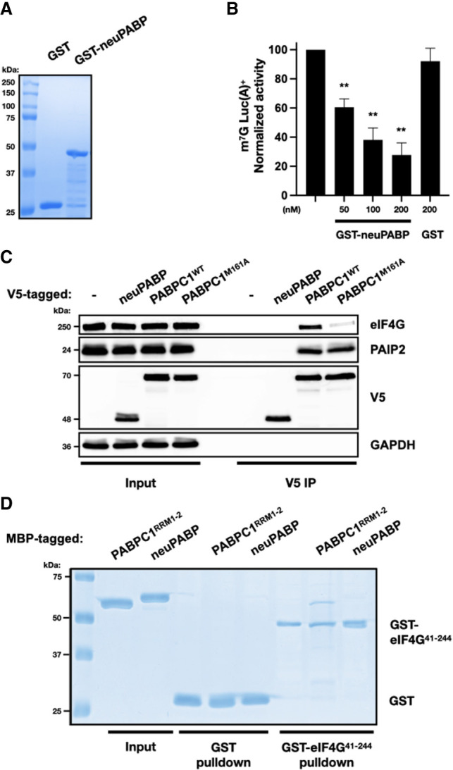
The mRNA 3' poly(A) tail plays a critical role in regulating both mRNA translation and turnover. It is bound by the cytoplasmic poly(A) binding protein (PABPC), an evolutionarily conserved protein that can interact with translation factors and mRNA decay machineries to regulate gene expression. Mammalian PABPC1, the prototypical PABPC, is expressed in most tissues and interacts with eukaryotic translation initiation factor 4G (eIF4G) to stimulate translation in specific contexts. In this study, we uncovered a new mammalian PABPC, which we named neural PABP (neuPABP), as it is predominantly expressed in the brain. neuPABP maintains a unique architecture as compared with other PABPCs, containing only two RNA recognition motifs (RRMs) and maintaining a unique N-terminal domain of unknown function. neuPABP expression is activated in neurons as they mature during synaptogenesis, where neuPABP localizes to the soma and postsynaptic densities. neuPABP interacts with the noncoding RNA BC1, as well as mRNAs coding for ribosomal and mitochondrial proteins. However, in contrast to PABPC1, neuPABP does not associate with actively translating mRNAs in the brain. In keeping with this, we show that neuPABP has evolved such that it does not bind eIF4G and as a result fails to support protein synthesis in vitro. Taken together, these results indicate that mammals have expanded their PABPC repertoire in the brain and propose that neuPABP may support the translational repression of select mRNAs.

Keywords: RNA binding protein; mRNA translation; poly(A) binding protein.


Figures

Figure 1.
Figure 1.
PABPC1L2 (neuPABP) displays a neural-specific expression pattern. (A) Schematic representation of PABPC1 and PABPC1L2 domain organization. (B) Schematic diagram of a human X chromosome showing the position of the Pabpc1l2 ampliconic gene. (C) Semiquantitative RT-PCR analysis of Pabpc1l2 and Actin mRNAs from multiple adult mouse tissues (C57BL/6J; age: 5 mo). (D) Western blotting of PABPC1, neuPABP, GAPDH, and Actin on lysates prepared from select adult mouse tissues (C57BL/6J; age: 5 mo).
Figure 2.
Figure 2.
neuPABP is a GUG-initiated protein with a misannotated N-terminal extension. (A) Schematic diagram of the predicted and revised neuPABP open reading frame, along with the predicted AUG and validated GUG initiator codons, respectively. The N-terminal region (highlighted in orange) corresponds to the domain of unknown function (DUF) that is predicted to be conserved between human, mouse, and rat neuPABP. (B) Schematic diagram of mouse neuPABP expression constructs containing C-terminal V5 tags. (C) Western blot analysis of HeLa cells transfected with plasmids encoding V5-tagged predicted neuPABP or containing the Pabpc1l2 5′ UTR containing the GTG codon or ATG codon.
Figure 3.
Figure 3.
neuPABP specificity for poly(A) RNA. (A) Summary of RNAcompete experiments for GST-neuPABP. The sequence logo of the neuPABP RNA binding motif is shown, along with a scatter plot displaying the Z scores and motifs for the two halves of the RNA pool (set A and set B). (B) Recombinant PABPC1 and neuPABP were analyzed by SDS-PAGE and Coomassie blue staining. (C) High-affinity binding of neuPABP to oligo(A) RNA. EMSA was carried out as described in the Materials and Methods. A constant amount of 32P-oligo(A)25 RNA was incubated with specific concentrations of neuPABP or PABPC1. The KD value of ∼50 nM was calculated from three biological experiments for both PABPC1 and neuPABP. Recombinant GST (control) did not lead to a gel shift of radiolabeled oligo.
Figure 4.
Figure 4.
neuPABP is expressed during neuronal maturation. (A) Subcellular fractionation of an adult mouse brain cortex (C57BL/6J; age: 2 mo) shows cytoplasmic localization of both neuPABP and PABPC1. GAPDH and hnRNPA1 were used as markers for cytoplasmic and nuclear fractions, respectively. (B) Western blot analysis of PABPC1, neuPABP, Actin, and β-tubulin III on lysates prepared from mouse brain cortices isolated at different stages of embryonic and postnatal development. (C) Western blot analysis of neuPABP, PSD-95, and β-tubulin III on lysates prepared from mouse primary cortical neurons. Neurons were isolated from P0 pups and cultured for defined days in vitro (DIV). (D) Western blot analysis of subcellular fractions of an adult mouse cortex (C57BL/6J; age: 6 mo) prepared by synaptosome fractionation. Lysates were probed with the postsynaptic (PSD) marker PSD-95 and the presynaptic marker synaptophysin (Syn), as well as neuPABP, PABPC1, and GAPDH. Cortex homogenates (H) were generated, and supernatant (S2) and the crude synaptosomal pellet (P2) were acquired after high-speed centrifugation of the S1 supernatant. The crude synaptosomal fraction was further fractionated into a Triton X-100-soluble non-PSD fraction (extrasynaptic) and a Triton X-100-insoluble PSD-containing fraction (synaptic).
Figure 5.
Figure 5.
neuPABP localizes with early RNP fractions on polysome gradients and interacts with specific RNAs. (A) Polysome profile traces of lysates prepared from mouse cortices (C57BL/6J; age: P9). (B) Lysates were fractionated by sucrose gradient centrifugation. Fractions were subsequently collected, TCA-precipitated, and resolved by SDS-PAGE, and Western blotting was subsequently performed using antibodies against neuPABP, PABPC1, and a ribosomal protein marker (RPS6). (C) Immunoprecipitation of neuPABP from an adult mouse hippocampus (C57BL/6J; age: 6 mo). Immunoprecipitated complexes were subjected to SDS-PAGE, and Western blotting was performed using anti-neuPABP and anti-GAPDH antibodies. neuPABP-enriched RNAs were isolated using RNA purification kit (Qiagen) and identified by RNA-seq. (D) Volcano scatter plot showing most significantly enriched RNAs with neuPABP (threshold set at log2 FC ≥ 1.5 and P-value set at <1 × 10⁻²⁰). BC1 RNA and mRNAs encoding ribosomal proteins (red) and nuclear-encoded mitochondrial proteins (blue) were enriched. (E) Top Wikipathway (WP) and associated gene ontology (GO) terms (cellular component) significantly enriched among proteins coded for by neuPABP-enriched mRNAs (FC ≥ 2). The number above each column represents the number of genes associated with its corresponding term. (F) RT-qPCR analyses of neuPABP-enriched transcripts identified by RNA-seq. neuPABP was immunoprecipitated from adult mouse hippocampi (C57BL/6J; age: 6 mo), and associated RNAs were Trizol-extracted. Error bars represent SEM from biological replicates (n = 3). Data points for biological replicates are shown as solid circles. Data were normalized to an in vitro transcribed RLuc spiked-in RNA.
Figure 6.
Figure 6.
neuPABP associates with untranslated mRNAs present in early RNP fraction. Cortices of adult mice (C57BL/6J; age: 6 mo) were triturated and formaldehyde-cross-linked. Lysates were prepared and fractionated by sucrose gradient centrifugation. (A) Ribosome traces of lysates prepared from formaldehyde-cross-linked adult mouse cortices (C57BL/6J; age: 6 mo). (B) Free RNP fractions (depleted of ribosomal subunits) were collected from the polysome gradient and resolved by SDS-PAGE, and Western blotting was performed using antibodies against RPS6, neuPABP, PABPC1, and GAPDH. (C) Immunoprecipitation of neuPABP from free RNP fractions from B. Immunoprecipitated complexes were resolved by SDS-PAGE, and Western blotting was performed using antibodies against neuPABP, PABPC1, and GAPDH. (D) RT-qPCR analysis of neuPABP-associated RNAs isolated from C. Error bars represent SEM from biological replicates (n = 3), which are shown as solid circles. A mitochondrial mRNA (mt.ND1) was used as a negative control. Data were normalized to an in vitro transcribed RLuc spiked-in RNA.
Figure 7.
Figure 7.
neuPABP represses translation in vitro and does not interact with eIF4G. (A) Recombinant GST and GST-tagged neuPABP were prepared and then analyzed by SDS-PAGE and Coomassie blue staining. (B) Capped poly(A)+ luciferase reporter RNA was incubated in Krebs-2 extract. Reactions were supplemented with either buffer alone (control), recombinant GST-neuPABP, or GST alone, as indicated. Normalized luciferase activity was measured relative to control. Error bars represent SEM from biological replicates (n = 3). A two-tailed Student's t-test (equal variance) was conducted (vs. control) to assess significance. (**) P-values <0.003 were calculated in GST-neuPABP treatment groups. (C) Immunoprecipitation of V5-tagged neuPABP, PABPC1WT, or PABPC1M161A from HeLa cells. Immunoprecipitated complexes were subjected to SDS-PAGE, and Western blot analysis was performed using anti-V5, anti-eIF4G, anti-PAIP2, and anti-GAPDH antibodies. (D) Recombinant glutathione-S-transferase (GST) or GST-tagged eIF4G41–244 was incubated with maltose-binding protein (MBP)-tagged PABPC1RRM1+2 or neuPABP. Precipitated proteins were separated by SDS-PAGE and visualized by Coomassie blue staining.
Figure 8.
Figure 8.
neuPABP has been selected to not bind eIF4G. (A) Schematic diagrams of PABPC1 and neuPABP domain organization, along with a comparative sequence analysis of human (Hs) and mouse (Mm) PABPC1 with human, mouse, and bat (Pk) neuPABP RRM2. Amino acids that play a role in PABPC1 binding to eIF4G are denoted by a green dot. Corresponding amino acids or those in proximity to eIF4G-intearcting residues are red. (B) Recombinant glutathione-S-transferase (GST)-tagged eIF4G41–244 was incubated with maltose-binding protein (MBP)-tagged PABPC1RRM1+2, neuPABPWT, or neuPABPMUT (Ile221Thr, Phe265Leu, and Tyr268Asp). Precipitated proteins were separated by SDS-PAGE and visualized by Coomassie staining. (C) Recombinant GST, GST-tagged neuPABPWT, and neuPABPMUT proteins were analyzed by SDS-PAGE and Coomassie blue staining. (D) Capped poly(A)+ luciferase reporter RNA was incubated in Krebs-2 extract. Reactions were supplemented with either recombinant GST, GST-neuPABPWT, or GST-neuPABPMUT, as indicated. Normalized luciferase activity was measured relative to buffer alone (control). Error bars represent SEM from biological replicates (n = 3). A two-tailed Student t-test (equal variance) was conducted (vs. GST) to access significance. (**) P-value <0.004 was calculated in GST-neuPABPWT treatment.
Figure 9.
Figure 9.
Model for the biological role of neuPABP. (Panel i) neuPABP binds to BC1 RNA and select translationally dormant mRNAs that may be transported to postsynaptic compartments. As neuPABP also lacks the PABPC1 MLLE domain, it may protect mRNAs from mRNA decay factors that can interact with this domain, including the PAN2–PAN3 complex and Tob, which interacts with the CCR4–NOT deadenylase complex. (Panel ii) It is possible that in specific contexts (depicted as a question mark), PABPC1 may displace neuPABP from mRNA poly(A) tails, bind eIF4G, and stimulate their mRNA translation.

References

    1. Aran D, Looney AP, Liu L, Wu E, Fong V, Hsu A, Chak S, Naikawadi RP, Wolters PJ, Abate AR, et al. 2019. Reference-based analysis of lung single-cell sequencing reveals a transitional profibrotic macrophage. Nat Immunol 20: 163–172. 10.1038/s41590-018-0276-y - DOI - PMC - PubMed
    1. Baer BW, Kornberg RD. 1980. Repeating structure of cytoplasmic poly(A)-ribonucleoprotein. Proc Natl Acad Sci 77: 1890–1892. 10.1073/pnas.77.4.1890 - DOI - PMC - PubMed
    1. Baer BW, Kornberg RD. 1983. The protein responsible for the repeating structure of cytoplasmic poly(A)-ribonucleoprotein. J Cell Biol 96: 717–721. 10.1083/jcb.96.3.717 - DOI - PMC - PubMed
    1. Biever A, Glock C, Tushev G, Ciirdaeva E, Dalmay T, Langer JD, Schuman EM. 2020. Monosomes actively translate synaptic mRNAs in neuronal processes. Science 367: eaay4991. 10.1126/science.aay4991 - DOI - PubMed
    1. Blanco P, Sargent CA, Boucher CA, Howell G, Ross M, Affara NA. 2001. A novel poly(A)-binding protein gene (PABPC5) maps to an X-specific subinterval in the Xq21.3/Yp11.2 homology block of the human sex chromosomes. Genomics 74: 1–11. 10.1006/geno.2001.6530 - DOI - PubMed

Publication types

Substances

LinkOut - more resources