. 2023 Sep 13;14(1):5660.
doi: 10.1038/s41467-023-41272-z.

De novo design of highly selective miniprotein inhibitors of integrins αvβ6 and αvβ8

Affiliations

De novo design of highly selective miniprotein inhibitors of integrins αvβ6 and αvβ8

Anindya Roy et al. Nat Commun. .

Abstract

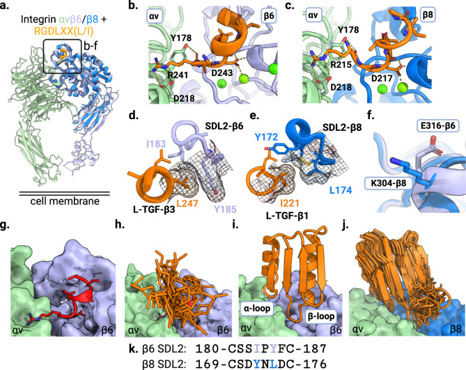
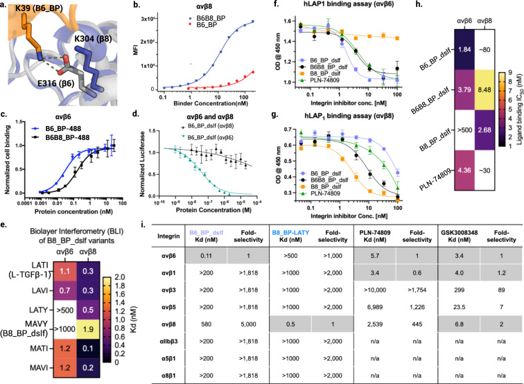
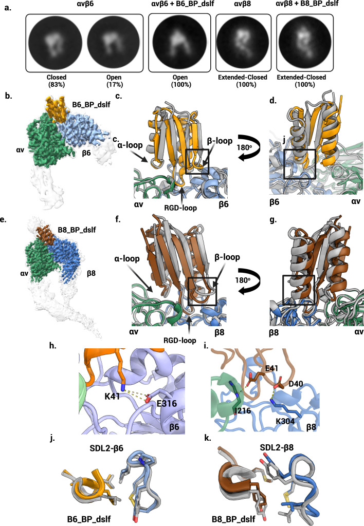
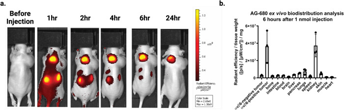
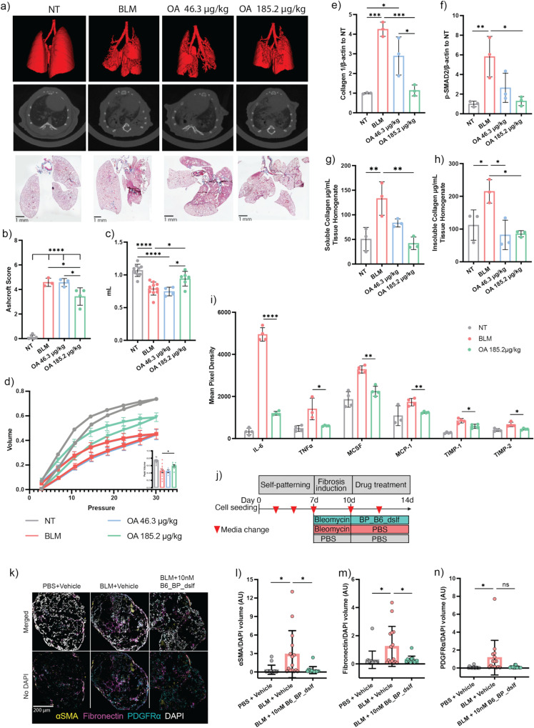
The RGD (Arg-Gly-Asp)-binding integrins αvβ6 and αvβ8 are clinically validated cancer and fibrosis targets of considerable therapeutic importance. Compounds that can discriminate between homologous αvβ6 and αvβ8 and other RGD integrins, stabilize specific conformational states, and have high thermal stability could have considerable therapeutic utility. Existing small molecule and antibody inhibitors do not have all these properties, and hence new approaches are needed. Here we describe a generalized method for computationally designing RGD-containing miniproteins selective for a single RGD integrin heterodimer and conformational state. We design hyperstable, selective αvβ6 and αvβ8 inhibitors that bind with picomolar affinity. CryoEM structures of the designed inhibitor-integrin complexes are very close to the computational design models, and show that the inhibitors stabilize specific conformational states of the αvβ6 and the αvβ8 integrins. In a lung fibrosis mouse model, the αvβ6 inhibitor potently reduced fibrotic burden and improved overall lung mechanics, demonstrating the therapeutic potential of de novo designed integrin binding proteins with high selectivity.


Conflict of interest statement

A.R., L.S, X.D., J.L. T.S., D.B. are co-inventors on an International patent (Serial # PCT/US2020/057016) filed by University of Washington covering molecules and their uses described in this manuscript. C. O. is an employee of AstraZeneca and may own stock or stock options. M.G.C is an inventor on “Antibodies that bind integrin avb8 and uses thereof”, U.S. Patent US20210277125A1. A.R., H.B., J.C.K., M.S., M.C. and D.B. are inventors on a provisional patent (patent application serial # 63/507,646) describing the sequence and usage of αvβ8 integrins binders. A.R., L.S., J.C.K, H.B. and D.B. are co-founders of Lila Biologics and own stock or stock options in the company. All other authors declare no competing interests.

Figures

Fig. 1
Fig. 1. Computational design of αvβ6 and αvβ8 selective minibinders.
a Crystal structure of β8 (PDB ID 6OM2) overlaid on the structure of αvβ6 integrin in complex with the L-TGF-β3 peptide RGDLXX(L/I) (PDB ID 4UM9). Inset highlights the zoomed-in regions shown in panels bf. The αv subunit is shown in green, β6 is in lavender, β8 is in blue, and the RGDLXX(L/I) peptide is in orange. b, c Shared polar interactions between the RGD motif and (b) αvβ6 and (c) αvβ8 integrin (L-TGF-β3/αvβ6 complex PDB ID 4UM9, L-TGF-β1/αvβ8 PDB ID 6OM2). d Hydrophobic packing of LXX(L/I) motif of L-TGF-β3 peptide with SDL2 of αvβ6 (PDB ID 4UM9). Leu247 packs optimally against Y185 from SDL2 of β6. e Hydrophobic packing of LXX(L/I) of L-TGF-β1 peptide with SDL2 of αvβ8 (PDB ID 6OM2). I221 of L-TGF-β1 peptide packs less tightly against L174 on SDL2 of β8 compared to the homologous interactions in panel d. f Charge reversal on β subunit: β8 contains K304 whereas the equivalent position on β6 is E316. g Surface structure of the αvβ6 integrin in complex with L-TGF-β3 peptide (red cartoon representation, PDB ID 4UM9). h Low RMSD matches to the L-TGF-β3 peptide bound to αvβ6 were harvested from the PDB database (orange stick representations). i Non-clashing fragments with αvβ6 were then incorporated in the α/β ferredoxin folds (orange ribbon representation) using Rosetta. j Loop extension strategy to design an αvβ8 selective minibinder: to make more extensive contacts to the β8 subunit the β-loop was resampled by one residue insertion (blue surface representation for β8 subunit, PDB ID 6OM2). In addition to the loop extension, the LXX(L/I) motif was allowed to be redesigned using Rosetta. k Partial sequence alignment of SDL2 of the β6/β8 subunits is shown highlighting two key positions packing against the LXX(L/I) motif of the L-TGF-β ligand (I183 and Y185 in SLD2-β6, and Y172 and L174 in SDL2-β8).
Fig. 2
Fig. 2. Selectivity of designed binders for αvβ6 and αvβ8.
a The A39K mutation confers selectivity towards αvβ6 compared to αvβ8 where there is a charge reversal (Glu316 for β6 shown as a gray stick, Lys304 for β8 shown as a blue stick). b Cell surface titration of B6B8_BP and B6_BP against K562 cells stably transfected with αvβ8. B6B8_BP lacking the A39K mutation binds to αvβ8 with a Kd of ~7.3 nM whereas B6_BP containing the A39K mutation binds to αvβ8 > 500 nM. c Cell surface titration of AlexaFluor-488-labeled B6B8_BP and B6_BP using αvβ6 (+) human epidermoid A431 carcinoma cells. B6_BP binds to A431 cells with higher potency than B6B8_BP (30 (±0.004) pM vs 167 (±0.028) pM). Data are presented as mean values +/− SD (n = 3 independent experiments). d B6_BP_dslf selectively inhibits αvβ6-mediated TGF-β1 activation. αvβ6 and αvβ8 transfectants were co-incubated with CAGA-reporter cells and GARP/TGF-β1 transfectants and inhibitors. B6_BP_dslf inhibits TGF-β activation with an IC50 of 32.8 (±3.4) nM. Data are presented as mean values +/− SD. e Binding affinities (Kd) of B8_BP_dslf (MAVY) point mutants to integrins αvβ6 and αvβ8, determined by BLI. Each experiment was repeated at least twice (n = 2). The LATI motif is in native L-TGF-β1. f, g Competitive inhibition of h-LAP1 binding to (f) αvβ6 and (g) αvβ8 by designed inhibitors and control small molecule PLN-74809. Each experiment was performed at least three times (n = 3). h Heatmap of IC50 values for h-LAP1 binding assays in f and g. i Binding affinities (Kd) and fold-selectivity values of B6_BP_dslf and B8_BP-LATY to all eight RGD integrins compared to small molecules PLN-74809 and GSK3008348. Binding data for PLN-74809 and GSK3008348 are taken from Decaris et al. 2021. Rows shaded in gray indicate the RGD integrin(s) for which each molecule is selective (i.e B6_BP_dslf and B8_BP-LATY are both mono-selective whereas PLN-74809 and GSK3008348 are dual- and tri-selective, respectively). n/a not available.
Fig. 3
Fig. 3. Structural characterization.
a Representative 2D class averages of integrin with and without minibinder. For αvβ6, both closed and open headpiece conformations are present in the unbound state, but in the presence of the minibinder the open conformation is dominant. For αvβ8, no open headpieces were observed, with or without minibinder. b CryoEM density map of αvβ6 bound to minibinder B6_BP_dslf. B6_BP_dslf (goldenrod) binds the integrin ligand binding cleft between the αv (green) and β6 (light blue) subunits and induces or stabilizes the open conformation. The sharpened, locally refined cryoEM map is shown in color, superimposed with the unsharpened map showing all domains of the αvβ6 headpiece in semi-transparent white. c, d Overlay of the designed αvβ6 + B6_BP_dslf model (gray) and the experimentally determined cryoEM model (colors). Although the overall angle of the minibinder is shifted, the RGD loop positioning is as predicted. Insets in c and d are magnified in panels h and i, respectively. e CryoEM density map of αvβ8 bound to minibinder B8_BP_dslf. Similar to B6_BP_dslf, B8_BP_dslf (brown) binds the integrin ligand binding cleft between the αv (green) and β8 (blue) subunits, however the conformation of αvβ8 remains in the closed headpiece conformation. The sharpened, locally refined cryoEM map is shown in color, superimposed with the unsharpened map showing all domains in the αvβ8 ectodomain construct in semi-transparent white. f, g An overlay of the designed αvβ8 + B8_BP_dslf model (gray) and the experimentally determined model. Although the overall angle of the minibinder is shifted, the RGD loop positioning is as predicted. Insets in f and g are magnified in panels i and k, respectively. h, i Key designed interactions between β-loop and β6/β8 subunit are observed in the cryoEM structure: K41 from B6_BP_dslf forms a salt bridge with E316 from the β6 subunit (panel h). E41 from β-loop makes backbone level hydrogen bond with I216 from β8 subunit and D40 makes salt bridge interaction with K304 from β8 subunit (panel i). j Experimental vs designed (gray) packing pattern of the LXXL motif and SDL2 of αvβ6. k Experimental vs designed (gray) packing pattern of the MAVY motif and SDL2 of αvβ8.
Fig. 4
Fig. 4. In vivo imaging of αvβ6 (+) A431 tumors using fluorescently labeled B6_BP.
a Athymic nude mice were injected with αvβ6 (+) A431 cells on the left shoulder and αvβ6 (−) HEK293T cells on the right shoulder. AlexaFluor-680-labeled B6_BP (AF680-B6_BP) was injected via the tail vein to image the tumors over time as indicated (see Supplementary for additional images, n = 5). b Semiquantitative ex vivo biodistribution assay of AF-680-B6_BP at 6 h post-tail vein injection. B6_BP selectively accumulates in αvβ6 (+) tumors and primarily clears via glomerular filtration in the kidneys. Data are presented as mean values +/− SD, n = 5 mice.
Fig. 5
Fig. 5. In vivo efficacy of OA-administered B6_BP_dslf in bleomycin-induced IPF.
a Three dimensional renderings of HR-uCT scans (top panel), representative HR-uCT scans (middle panel), and representative Masson-trichrome images for nontreated (NT), bleomycin treated (BLM) and inhaled B6_BP_dslf groups. b Average Ashcroft Scoring of Masson-trichrome images (data represented as mean ± SEM, Tukey’s t-test, NT vs BLM P value = <0.0001, NT vs B6_BP_dslf 46.3 ug/kg P value = <0.0001, NT vs B6_BP_dslf 185.2 ug/kg P value = <0.0001, BLM vs B6_BP_dslf 185.2 ug/kg P value = 0.0214, B6_BP_dslf 46.3 ug/kg vs B6_BP_dslf 185.2 ug/kg P value = 0.0232). c Forced vital capacity as measured by SCIREQ flexiVent FX (data represented as mean ± SEM, Tukey’s t-test, NT vs BLM P value = <0.0001, NT vs B6_BP_dslf 46.3 ug/kg P value = <0.0001, BLM vs B6_BP_dslf 185.2 ug/kg P value = 0.034, B6_BP_dslf 46.3ug/kg vs B6_BP_dslf 185.2 ug/kg P value = 0.0386). d Pressure-Volume curves measured by SCIREQ flexiVent with peak volumes in the inset graph (data represented as mean ± SEM, Tukey’s t-test, BLM vs B6_BP_dslf 185.2 ug/kg P value = 0.0407). e Whole lung tissue homogenate western blot analysis of Collagen1 (data represented as mean ± SD, Tukey’s t-test, NT vs BLM P value = 0.0003, NT vs B6_BP_dslf 46.3 ug/kg P value = 0.0109, BLM vs B6_BP_dslf 185.2 ug/kg P value = 0.0005, B6_BP_dslf 46.3 ug/kg vs B6_BP_dslf 185.2 ug/kg P value = 0.0165) and f p-SMAD2 (data represented as mean ± SD, Tukey’s t-test, NT vs BLM P value = 0.0082, BLM vs B6_BP_dslf 185.2 ug/kg P value = 0.0117) show a dose-dependent reduction of these pro-fibrotic markers following B6_BP_dslf OA treatment. g Soluble (data represented as mean ± SD, Tukey’s t-test, NT vs BLM P value = 0.0078, BLM vs B6_BP_dslf 185.2 ug/kg P value = 0.0043) and h Insoluble (data represented as mean ± SD, Tukey’s t-test, NT vs BLM P value = 0.0382, BLM vs B6_BP_dslf 46.3 ug/kg P value = 0.01, BLM vs B6_BP_dslf 185.2 ug/kg P value = 0.0117) collagen levels are lower following B6_BP_dslf OA treatment. i Cytokine array analysis of common cytokines implicated in inflammation and IPF (all data represented as mean ± SD, all NT vs. BLM are significant, p-value < 0.05). j Time course of hFLO organoid growth, bleomycin induction, and 10 nM B6_BP_dslf treatment. k Fluorescent confocal microscopy imaging of hFLO sections immunostained with pro-fibrotic markers αSMA, fibronectin, and PDGFRα. (ln are presented as bar graphs with mean ± SD, N = 15 cell aggregates were used per treatment, *P < 0.05 determined using two-tailed Welch’s t-test, box and whiskers show the mean and the maximum and minimum values) Volumetric analysis of (l) αSMA (PBS vs BLM P value = 0.0238, BLM vs BLM + 10 nM B6_BP_dslf P value = 0.0205) (m) fibronectin (PBS vs BLM P value = 0.0285, BLM vs BLM + 10 nM B6_BP_dslf P value = 0.0242, and n PDGFRα (PBS vs BLM P value = 0.0451) normalized to DAPI signal. *p-value < 0.05, **p-value < 0.01, ***p-value < 0.001, ****p-value < 0.0001.

Update of

Similar articles

Cited by

References

    1. Dong X, Hudson NE, Lu C, Springer TA. Structural determinants of integrin β-subunit specificity for latent TGF-β. Nat. Struct. Mol. Biol. 2014;21:1091–1096. doi: 10.1038/nsmb.2905. - DOI - PMC - PubMed
    1. Campbell MG, et al. Cryo-EM Reveals Integrin-Mediated TGF-β Activation without Release from Latent TGF-β. Cell. 2020;180:490–501.e16. doi: 10.1016/j.cell.2019.12.030. - DOI - PMC - PubMed
    1. Dong X, et al. Force interacts with macromolecular structure in activation of TGF-β. Nature. 2017;542:55–59. doi: 10.1038/nature21035. - DOI - PMC - PubMed
    1. Horan GS, et al. Partial inhibition of integrin alpha(v)beta6 prevents pulmonary fibrosis without exacerbating inflammation. Am. J. Respir. Crit. Care Med. 2008;177:56–65. doi: 10.1164/rccm.200706-805OC. - DOI - PubMed
    1. Saini G, et al. αvβ6 integrin may be a potential prognostic biomarker in interstitial lung disease. Eur. Respir. J. 2015;46:486–494. doi: 10.1183/09031936.00210414. - DOI - PubMed

Publication types