Review
. 2023 Sep 14;80(10):291.
doi: 10.1007/s00018-023-04914-5.

Revolutionizing control strategies against Mycobacterium tuberculosis infection through selected targeting of lipid metabolism

Affiliations
Review

Revolutionizing control strategies against Mycobacterium tuberculosis infection through selected targeting of lipid metabolism

Hagyu Kim et al. Cell Mol Life Sci. .

Abstract

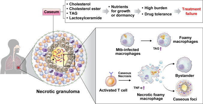
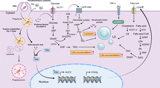
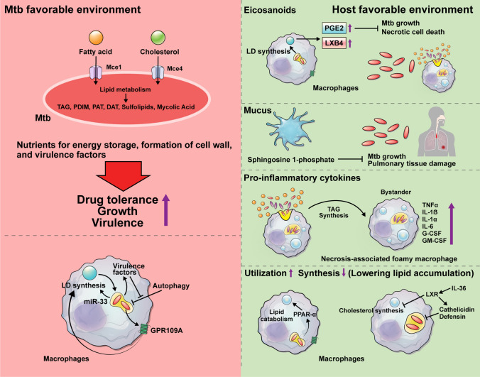
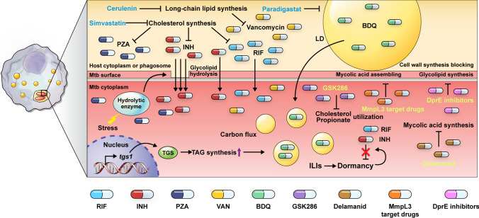
Lipid species play a critical role in the growth and virulence expression of Mycobacterium tuberculosis (Mtb), the causative agent of tuberculosis (TB). During Mtb infection, foamy macrophages accumulate lipids in granulomas, providing metabolic adaptation and survival strategies for Mtb against multiple stresses. Host-derived lipid species, including triacylglycerol and cholesterol, can also contribute to the development of drug-tolerant Mtb, leading to reduced efficacy of antibiotics targeting the bacterial cell wall or transcription. Transcriptional and metabolic analyses indicate that lipid metabolism-associated factors of Mtb are highly regulated by antibiotics and ultimately affect treatment outcomes. Despite the well-known association between major antibiotics and lipid metabolites in TB treatment, a comprehensive understanding of how altered lipid metabolites in both host and Mtb influence treatment outcomes in a drug-specific manner is necessary to overcome drug tolerance. The current review explores the controversies and correlations between lipids and drug efficacy in various Mtb infection models and proposes novel approaches to enhance the efficacy of anti-TB drugs. Moreover, the review provides insights into the efficacious control of Mtb infection by elucidating the impact of lipids on drug efficacy. This review aims to improve the effectiveness of current anti-TB drugs and facilitate the development of innovative therapeutic strategies against Mtb infection by making reverse use of Mtb-favoring lipid species.

Keywords: Anti-TB drugs; Drug efficacy; Drug tolerance; Lipid droplet; Lipid metabolism; Mycobacterium tuberculosis.


Conflict of interest statement

The authors declare that the research was conducted in the absence of any commercial or financial relationships that could be construed as a potential conflict of interest.

Figures

Fig. 1
Fig. 1
Caseum development is the process of lipid enrichment with necrosis, leading to cause treatment failure. Following the formation of granuloma against Mtb infection, necrotized foamy macrophages that surround the caseous center gradually begin to form the lipid-rich environment of the caseum. Caseum includes high TAG, cholesterol, lactosylceramide, and cholesterol ester. These lipid species are utilized for the replication of Mtb as well as induce a dormancy state that decreases drug efficacy. In the granuloma, Mtb-infected macrophages become lipid-rich foamy macrophages. Moreover, caseous necrosis with the necrotic death of infected foamy macrophages by activated T cells produces interferons and activates adjacent macrophages to lead to caseous necrosis. In addition, TAG-rich foamy macrophages are present in necrotic granulomas, and the presence of these macrophages is associated with a high level of TNF-α in the necrotic core. Take together acceleration of necrotic cell death in caseous foci provides nutrients for replication and confers TB drug tolerance to Mtb, suggesting inhibition of mycobacterial clearance, resulting in treatment failure. Mtb Mycobacterium tuberculosis, TAG triacylglyceride, TNF-α tumor necrosis factor-α
Fig. 2
Fig. 2
Mechanisms of accumulation of lipid droplets induced by Mtb in infected host cells. Through phagocytosis, macrophages take Mtb into the cytosol. One of the events following Mtb infection is modulatory effects on the lipid metabolisms in the macrophages, causing foamy macrophages. ESAT-6 activates the lipolysis inhibitory receptor, GPR109A by increasing glucose imports with perturbation of glycolysis and formation of ketone body, 3HB. The presence of ESAT-6 causes the glycerol level to rise as a result of the reversible conversion of DHAP by GPDH, producing assessed de novo TAG production. Additionally, the oxygenated mycolic acid, keto-mycolic acid induces the ability to transactivate TR4 and produces foamy macrophages. Increased lipid uptake by scavenger receptor CD36 is induced by PPARg and Nr2C2, which encode PPAR-γ and TR4, respectively. Additionally, Mtb alters the signaling pathways of nuclear transcription factors. For instance, TNF receptor signaling is induced by the infection of macrophages through downstream activation of the caspase cascade and mTORC1, which leads to the accumulation of LD as a result of decreased fatty acid consumption. 1-TbAd, a terpenyl nucleoside released from Mtb, is responsible for inducing lipid accumulation in the phagolysosome of macrophages. Mtb Mycobacterium tuberculosis, ESAT-6 6-kDa early secretory antigenic, LDs lipid droplets, TR4 testicular receptor 4 nuclear receptor, TNF tumor necrosis factor, 1-TbAd 1-tuberculosinyladenosine, 3HB 3-hydroxy butyrate, G6P glucose-6- phosphate, DHAP dihydroxy acetone phosphate, GPDH glycerol-3-phosphate dehydrogenase, G3P Glycerol 3-phosphate, DAG diacylglycerol, TAG triacylglyceride, 3PG 3-phosphoglycerate, 2PG 2-phosphoglycerate, PEP phosphoenol pyruvate, PKA protein kinase A, HSL hormone-sensitive lipase, ACS acyl-CoA synthetase, PA phosphatidic acid, GPAT glycerol-3-phosphate O-acyltransferase, LPA lysophosphatidic acid, AGPAT 1-acylglycerol-3-phosphate O-acyltransferase, PAP phosphatidic acid phosphatase, DGAT diacylglycerol O-acyltransferases, ACAT acyl-CoA: cholesterol acyltransferase, CE cholesterol ester.
Fig. 3
Fig. 3
Lipid metabolism favoring for Mycobacterium tuberculosis (left side) and host (right side). Lipids in the host environment are related to various roles during Mtb infection. Mtb utilizes fatty acids as its major source of energy rather than carbohydrates. As lipid transporters, Mce1 and Mce4 are required necessarily for the import of fatty acids and cholesterol respectively. Following the influx of fatty acid and cholesterol into Mtb, those lipids are exploited as sources of lipid metabolism, which generates several vital metabolites for persistence and survival, including TAG, PAT, DAT, mycolic acid, and PDIM. The metabolites confer drug tolerance, capacity of replication, and virulence to Mtb, suggesting that lipids from the host are beneficial for Mtb infection. miR-33 generated by Mtb and the miR-33 passenger strand reduce mitochondrial FAO and increase LD formation in macrophages. In contrast, several responses involved in lipids contribute to host defense against Mtb infection. PGE2 and LXB4 are two host-protective arachidonic acid-derived eicosanoids whose production is specifically boosted by LD formation during Mtb infection. One of the components of mucus, S1P suppresses mycobacterial growth and tissue damage. In addition, increased TAG synthesis in necrosis-associated foamy macrophage induces the expression of pro-inflammatory cytokines such as TNF-α, IL-1β, IL-1α, IL-6, and G-CSF/GM-CSF. PPAR-α, a member of another PPAR family, is activated by Mtb to increase fatty acid β-oxidation and lipid catabolism in the host cell. Infected macrophages that have PPAR-α activation elevate the expression of genes related to FAO. Using IL-36 signaling, the LXR is a master regulator of cholesterol. By inhibiting the synthesis of intracellular cholesterol, induction of LXR activation reduces the growth of Mtb. It also regulates production of antimicrobial peptides including cathelicidin and defensin. Mtb Mycobacterium tuberculosis, LD lipid droplet, TAG triacylglyceride, PAT polyacyltrehalose, DAT diacyltrehaloses, PDIM phthiocerol dimycocerosate, miR microRNA, PGE2 prostaglandin E2, LXB4 lipoxin B4, S1P sphingosine 1-phospate, TNF-α tumor necrosis factor-α, IL- interleukin-, G-CSF/GM-CSF granulocyte colony-stimulating factor /granulocyte–macrophage colony-stimulating factor, FAO fatty acid oxidation, LXR liver X receptor
Fig. 4
Fig. 4
STRING data analysis of lipid metabolic genes in Mtb following anti-TB drugs administration. STRING analysis (http://string-db.org/) reveals the putative interactions of lipid metabolism-involved genes in Mtb by drug exposure. Red color means upregulated expression and blue color means downregulated expression by the response to antibiotics. All lines of data represent the active interaction sources of experiments, co-expression, neighborhood, gene fusion, and co-occurrence. R rifampin, H isoniazid, E ethambutol, Z pyrazinamide, VAN vancomycin.
Fig. 5
Fig. 5
Various mechanisms related to drug efficacy and lipid metabolism during Mtb infection. Lipid metabolic switching in Mtb results in a dormancy-like phenotype as well as persistence causes tolerance of bactericidal drugs. tgs1, encoding the gene of triacylglycerol synthase, induces the formation of ILI in Mtb. Since the accumulation of lipids in the ILI of mycobacteria confers dormancy- like phenotype in Mtb-infected foamy macrophage, anti-TB antibiotics such as RIF and INH exhibit low efficacy. It is crucial for antibiotic tolerance that carbon fluxes are modulated to limit TAG production, which is brought on by various stress responses during Mtb infection. Adjunctive therapy with statins such as simvastatin, a lipid-lowering drug by inhibition of cholesterol synthetic pathway, enhance the efficacy of combination with RIF, INH, and PZA. In addition, another adjunctive component, cerulenin, a potent long-chain lipid synthesis inhibitor, increases the susceptibility of VAN to clinical isolates. Recently, a study demonstrated the possibility that lipids could improve medication efficacy. BDQ is a lipophilic drug and is absorbed in LD in host cells. Interestingly, Mtb consumes nutrients from the LD with BDQ, indicating that lipids promote the efficacy of BDQ. Furthermore, a LD inhibitor, paradigastat reduces the BDQ abundance in Mtb. Additionally, an enzyme that hydrolyzes the mycobacterial glycolipid TDM, reforms the Mtb cell wall to increase nutrient influx as well as sensitize to INH under the stress. GSK286 is an inhibitor of cholesterol propionate catabolism. Delamanid inhibits mycolic acid synthesis through mycobacterial F420 system. MmpL3 target drugs such as SQ109 and BM212 prevent mycolic acid assembling for mycobacterial cell wall formation. DprE inhibitors like benzothiazinone impede cell wall synthesis related to the formation of lipoarabinomannan and arabinogalactan. Mtb Mycobacterium tuberculosis, LD lipid droplet, TAG triacylglyceride, ILI intracytoplasmic lipid inclusion, TDM trehalose-6, 6-dimycolate, RIF rifampin, INH isoniazid, PZA pyraziamide, BDQ bedaquiline, VAN vancomycin, Tsg1 triacylglycerol synthase 1, MmpL3 Mycobacterial membrane protein large 3

Similar articles

Cited by

References

    1. World Health O . Global tuberculosis report 2022. Geneva: World Health Organization; 2022.
    1. Russell DG, Cardona PJ, Kim MJ, et al. Foamy macrophages and the progression of the human tuberculosis granuloma. Nat Immunol. 2009;10(9):943–948. doi: 10.1038/ni.1781. - DOI - PMC - PubMed
    1. Datta M, Via LE, Kamoun WS, et al. Anti-vascular endothelial growth factor treatment normalizes tuberculosis granuloma vasculature and improves small molecule delivery. Proc Natl Acad Sci USA. 2015;112(6):1827–1832. doi: 10.1073/pnas.1424563112. - DOI - PMC - PubMed
    1. Ramakrishnan L. Revisiting the role of the granuloma in tuberculosis. Nat Rev Immunol. 2012;12(5):352–366. doi: 10.1038/nri3211. - DOI - PubMed
    1. Sarathy JP, Dartois V. Caseum: a Niche for Mycobacterium tuberculosis drug-tolerant persisters. Clin Microbiol Rev. 2020 doi: 10.1128/cmr.00159-19. - DOI - PMC - PubMed