Review
. 2023 Sep 13;18(1):103.
doi: 10.1186/s13000-023-01390-0.

Extraskeletal myxoid chondrosarcoma of the gingival: a rare case report and review of the literature

Affiliations
Review

Extraskeletal myxoid chondrosarcoma of the gingival: a rare case report and review of the literature

Jiaqi Li et al. Diagn Pathol. .

Abstract

Background: Extraskeletal myxoid chondrosarcoma (EMC) is a rare malignant tumor described in the head and neck region, especially in the gingival. We present one case arising in the gingival of right mandible, and briefly reviewed the related literature.

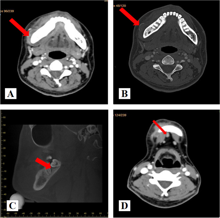
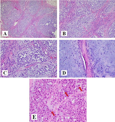
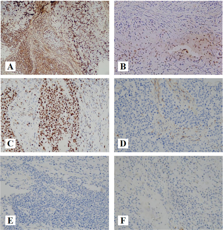
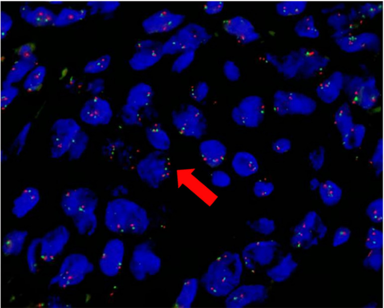
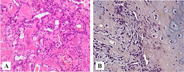
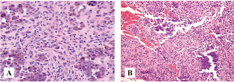
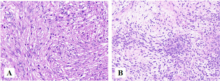
Case presentation: A 24-year-old male patient with a lesion of 3.5*2.0 cm in buccal gingival of right posterior mandible for 2 months. The tumor was composed of cartilaginous structures and myxoid matrix. Immunohistochemical(IHC) showed that the tumor cells to be positive for vimentin, focally positive for S-100, negative for calponin, SMA, SOX10. The Ki-67 labelling index was 80%. Fluorescent in situ Hybridization (FISH) was positive for NR4A3 rearrangement.

Conclusions: Due to its unusual site and low incidence in the oral region, a combination of histological findings, immunohistochemistry, and molecular pathology as well as differential diagnosis with other diseases should be taken into consideration in the process of clinical diagnosis and treatment.

Keywords: Differential diagnoses; Extraskeletal myxoid chondrosarcoma; Immunohistochemistry; Malignant tumor; Mandibular gingiva; NR4A3.


Conflict of interest statement

The authors declare no competing interests.

Figures

Fig. 1
Fig. 1
Imaging manifestations of EMC. A soft tissues of the labial buccal and lingual side of the right mandible were irregularly thickened(arrow). B There appeared to be irregular periosteal reaction in the lateral part of the right mandible(arrow). C periodontal space broading were observed in the 47 distal and 48 teeth(arrow).(D)There was no obvious enlargement of submental lymph nodes(arrow)
Fig. 2
Fig. 2
H&E findings of EMC. A (HE × 40): the tumor cells immersed in a myxoid stroma in a multilobular arrangement, which is separated by fibrous septae. B (HE × 100): The cells were arranged in ribbons, trabeculae and small nests present in a myxoid background. C (HE × 200): At high magnification, the tumor cells were uniform in size, with round and oval nuclei, deep chromatin and large nuclear atypia. D (HE × 200): The tumor cells were located in the cartilage lacunae, showing atypia and mitosis. E (HE × 400): Mitosis was obviously(arrow)
Fig. 3
Fig. 3
The immunoprofile of EMC. A (IHC × 100): Vimentin showing positive immunoreactivity in the cytoplasm. B (IHC × 200): S-100 showing focally positive immunoreactivity in the cytoplasm and nucleus. C (IHC × 200): Nuclear staining for Ki-67 with index labeling of 80%. D (IHC × 200): Smooth muscle actin (SMA), negative. E (IHC × 200): KRT7, negative. F (IHC × 200): SOX10, negative
Fig. 4
Fig. 4
FISH images of the tumor. Orange (R) signal marks the 3 'end of NR4A3 gene, green (G) signal marks the 5 'end of NR4A3 gene. The normal signal mode is 2F (F is the yellow signal of red-green fusion), and the typical positive signal mode is 1F1R1G. FISH analysis on the histological section evidenced rearrangement of NR4A3 gene(arrow)
Fig. 5
Fig. 5
Epidemiological findings of EMC. A Male–female ratio of EMC patients. B Age distribution of EMC patients. C The distribution of the primary lesion sites in the head and neck region
Fig. 6
Fig. 6
A (HE × 40): Lobules of uniform spindle or stellate cells with myxoid or chondroid to fibrous stroma of CMF. B (IHC × 200): CD10, positive [17, 18]
Fig. 7
Fig. 7
A (HE × 40): stellate and spindle-shaped cells embedded in a richly myxoid extracellular matrix. B (HE × 200): Residual odontogenic epithelium can be seen(arrow)
Fig. 8
Fig. 8
A (HE × 400): Abundant myxoid stroma, proliferation of lipoblasts in variable numbers and fine vascularization, A network of chicken's foot like capillaries can be seen in the mesenchyma. B Cytogenetic and molecular cytogenetic findings in a myxoid liposarcoma with cryptic EWSR1-DDIT3 fusion(arrow). Metaphase fluorescence in situ hybridization (FISH) with a break-apart probe specific for the EWSR1 gene. An intact yellow signal is seen on the normal chromosome 22, whereas a split signal is seen on the derivative chromosomes 11 (green) and 22 (red) [26, 27]
Fig. 9
Fig. 9
A (HE × 40): Architecturally, the tumor cells are arranged into nests and a large number of mucus-like areas can be seen. B (HE × 100): The tumor cells are plasmacytoid cells
Fig. 10
Fig. 10
A Interconnecting trabeculae of woven bone rimmed by plump osteoblasts and richly vascularized fibroblastic stroma are present. B Substantial chondroblastic differentiation and nuclear atypia [33, 34]
Fig. 11
Fig. 11
A (HE × 40): Chondrocytes with deposition of hemosiderin deposition. B (HE × 40): The calcifications present as ‘‘chicken-wire’’ appearance [37]
Fig. 12
Fig. 12
A Monophasic SS, made of spindle cells with moderate cytologic atypia and differentiated areas of variable cellularity. B Biphasic SS composed by glandular-like structures with spindle cells [38]

References

    1. Romañach MJ, Carlos R, Nuyens M, de Andrade BA, de Almeida OP. Extraskeletal myxoid chondrosarcoma of the masticator space in a pediatric patient. J Clin Exp Dent. 2017;9(6):e825–e831. - PMC - PubMed
    1. Garde JB, Palaskar SJ, Kathuriya PT. Extraskeletal myxoid chondrosarcoma of maxilla: A rare entity. J Oral Maxillofac Pathol. 2016;20(1):151–3. doi: 10.4103/0973-029X.180980. - DOI - PMC - PubMed
    1. Goh YW, Spagnolo DV, Platten M, Caterina P, Fisher C, Oliveira AM, Nascimento AG. Extraskeletal myxoid chondrosarcoma: a light microscopic, immuno-histochemical, ultrastructural and immuno-ultrastructural study indicating neuroendocrine differentiation. Histopathology. 2001;39(5):514–524. doi: 10.1046/j.1365-2559.2001.01277.x. - DOI - PubMed
    1. Brenca M, Stacchiotti S, Fassetta K, Sbaraglia M, Janjusevic M, Racanelli D, Polano M, Rossi S, Brich S, Dagrada GP, Collini P, Colombo C, Gronchi A, Astolfi A, Indio V, Pantaleo MA, Picci P, Casali PG, Dei Tos AP, Pilotti S, Maestro R. NR4A3 fusion proteins trigger an axon guidance switch that marks the difference between EWSR1 and TAF15 translocated extraskeletal myxoid chondrosarcomas. J Pathol. 2019;249(1):90–101. doi: 10.1002/path.5284. - DOI - PMC - PubMed
    1. Chen Y, Zhang Y. Myxoid chondrosarcoma of the mandible in a 22-year-old man: a case report. Mol Clin Oncol. 2016;5(3):237–240. doi: 10.3892/mco.2016.939. - DOI - PMC - PubMed

Supplementary concepts

LinkOut - more resources