

Case Reports
doi: 10.1002/ccr3.7894.
eCollection 2023 Sep.
Black discoloration of the knee articular cartilage in a patient with pigmented villonodular synovitis: A case report
Affiliations
- PMID: 37705584
- PMCID: PMC10495615
- DOI: 10.1002/ccr3.7894
Item in Clipboard
Case Reports
Black discoloration of the knee articular cartilage in a patient with pigmented villonodular synovitis: A case report
Clin Case Rep.
.
Abstract
In this case report, total knee arthroplasty was performed in a patient with pigmented villonodular synovitis. During surgery, severe black discoloration of the articular cartilage and menisci was observed in the patient. According to literatures, this is the first case report of severe articular cartilage pigmentation in a patient with pigmented villonodular synovitis.
Keywords: PVNS; black cartilage; black menisci; case report; total knee arthroplasty.
© 2023 The Authors. Clinical Case Reports published by John Wiley & Sons Ltd.
Conflict of interest statement
The authors declare that they have no conflict of interests.
Figures

Hip‐knee‐ankle radiographic with joint space narrowing, subchondral sclerosis, and osteophyte formation.
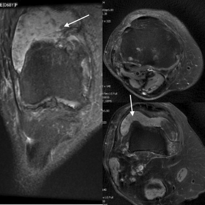
Knee joint effusion and popliteal cyst with internal debris which located posteriorly, suggesting the possibility of pigmented villonodular synovitis (PVNS).

Severe black pigmentation of the synovial membrane and menisci and pigmentation of the articular surface.

Postsurgical radiographic with acceptable alignment.

The lesion is composed of papillary, villous, nodular, and pseudoglandular or cleft like spaces with a synovial lining. Osteoclast‐like multinucleated giant cells and hemosiderin‐laden macrophages are seen. Histopathologic evaluation is compatible with pigmented villonodular synovitis (PVNS).
References
-
- Raaijmaakers M, Steenbrugge F, Dierickx C. Ochronosis, arthroscopy of a black knee: a case report and review of the literature. Knee Surg Sports Traumatol Arthrosc. 2008;16(2):182‐184. - PubMed
-
- Sudesh G. Pigmentation of articular surfaces of bones as an identification factor: a unique case report. Am J Forensic Med Pathol. 2006;27:161‐162. - PubMed
-
- Lee CA, Kessler CM, Varon D, et al. Synovium in haemophilic arthropathy. Haemophilia. 1998;4(4):502‐505. - PubMed
-
- Connolly CE, O'Reilly U, Donlon J. Black cartilage associated with levodopa. The Lancet. 1986;327(8482):690. - PubMed
Publication types
LinkOut - more resources
Full Text Sources
