. 2023 Aug 28:21:4207-4214.
doi: 10.1016/j.csbj.2023.08.027. eCollection 2023.

Variation of extrachromosomal circular DNA in cancer cell lines

Affiliations

Variation of extrachromosomal circular DNA in cancer cell lines

Carl Rung Dos Santos et al. Comput Struct Biotechnol J. .

Abstract

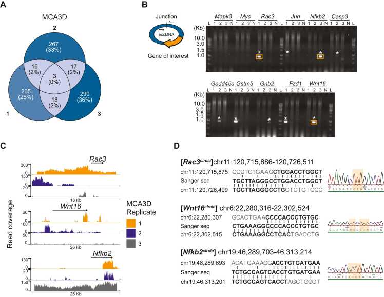
The presence of oncogene carrying eccDNAs is strongly associated with carcinogenesis and poor patient survival. Tumour biopsies and in vitro cancer cell lines are frequently utilized as models to investigate the role of eccDNA in cancer. However, eccDNAs are often lost during the in vitro growth of cancer cell lines, questioning the reproducibility of studies utilizing cancer cell line models. Here, we conducted a comprehensive analysis of eccDNA variability in seven cancer cell lines (MCA3D, PDV, HaCa4, CarC, MIA-PaCa-2, AsPC-1, and PC-3). We compared the content of unique eccDNAs between triplicates of each cell line and found that the number of unique eccDNA is specific to each cell line, while the eccDNA sequence content varied greatly among triplicates (∼ 0-1% eccDNA coordinate commonality). In the PC-3 cell line, we found that the large eccDNA (ecDNA) with MYC is present in high-copy number in an NCI cell line isolate but not present in ATCC isolates. Together, these results reveal that the sequence content of eccDNA is highly variable in cancer cell lines. This highlights the importance of testing cancer cell lines before use, and to enrich for subclones in cell lines with the desired eccDNA to get relatively pure population for studying the role of eccDNA in cancer.

Keywords: CNV; Cancer cell lines; Double minute; EcDNA; EccDNA; Reproducibility; non-mendelian.


Conflict of interest statement

B.R. is cofounder of CARE-DNA. All other authors declare that they have no known competing financial interests or personal relationships that could have appeared to influence the work reported in this paper.

Figures

ga1
Graphical abstract
Fig. 1
Fig. 1
Workflow and summary of eccDNA extracted with Circle-Pure identified with Circle-Map. A) Schematic representation of the study workflow. B) Mean number of unique eccDNA (n = 3) identified by Circle-Map. The presence of all eccDNAs reported in the mouse cancer cell lines is supported by at least 1 soft-clipped read, 1 discordant read-pair, and at least 90% read coverage to be considered valid. The presence of all eccDNAs reported in human cancer cell lines is supported by at least 4 soft-clipped reads, 4 discordant read-pairs, and at least 99% read coverage to be considered valid. C) Saturation plot of relative read count percentage against the eccDNA count of the cell line replicates with highest eccDNA count/million mapped reads. D) A PFGE gel of total DNA extracted from MIA-PaCa-2 (rep 3) and AsPC-1 (rep 3) with Circle-Pure. L = CHEF DNA Size Marker, 0.2–2.2 Mb, S. cerevisiae Ladder.
Fig. 2
Fig. 2
Inter-replicate eccDNA variation. A) Venn diagram of unique full-length protein-coding genes located on eccDNA in each replicate (designated 1, 2, and 3) of the MCA3D cell line. Full-length protein coding genes located on eccDNA found in three replicate: Krtap10–4, Gm10840, Psmg3. Of the total number of unique eccDNAs identified in the MCA3D cell line, between ∼ 0.2–0.5% carry a full-length protein coding gene. B) Outwards PCR spanning the junction site of 11 eccDNAs identified in MCA3D that carry a known oncogene. Amplified eccDNA was used as the template DNA for the PCR reaction. The expected band size ranged from 0.8 kb to 1.2 kb. L = GeneRuler 1Kb DNA ladder. N = negative control. Lane numbers refer to replicate numbers. Stars indicate correct size of PCR product and orange boxes refer to the gel slices that were extracted for Sanger sequencing. C) eccDNA read coverage plot of Rac3circle, Wnt16circle and Nfkb2circle. D) Sanger sequencing results of the PCR bands highlighted in orange in B.
Fig. 3
Fig. 3
Copy-number variation and eccDNA identification in PC-3. A) Venn diagram of genes located on amplified eccDNAs (copy number > 5) in PC-3 cells from this study (left), comparison with Seim et al. (middle), and comparison with Turner et al. (Right). B) CNVkit scatterplot of chr8:120–140 Mb of the PC-3 cell line isolate from this study (left), Seim et al. (middle), and Turner et al. (Right). The y-axis describes the log(2) copy ratio reported by CNVkit and represents the deviation in copy number of each genomic segment in the sample relative to the expected copy number based on the reference genome . The vertical purple line represents the location of the MYC gene (∼127 Mb). C) AmpliconArchitect output of the amplicon that included the MYC gene in the PC-3 cell line isolate from this study (left), Seim et al. (Middle), and Turner et al. (Right). Read coverage is represented as grey coverage bars and absolute copy-number is represented as horizontal orange lines. The location of the MYC and PVT1 gene is shown as purple lines underneath the graphs. D) No MYC-carrying eccDNAs were identified in the PC-3 cell line isolate from this study (left) or with data from Seim et al. (middle). With the Turner et al. WGS data, MYC and PVT1 carrying eccDNAs were identified by AmpliconClassifier and visualized with CycleViz (right).
Fig. 4
Fig. 4
MYC gene copy number in PC-3 cells by FISH analysis. A) Representative FISH metaphase chromosomes in PC-3 cells with MYC (red) and stained with DAPI (cyan) for DNA detection. B) Number of MYC copies detected in 74 PC-3 metaphase chromosome spreads. Median number of MYC copies per cell = 6.

References

    1. Gaubatz J.W. Extrachromosomal circular DNAs and genomic sequence plasticity in eukaryotic cells. Mutat Res DNAging. 1990;237:271–292. - PubMed
    1. Møller H.D., et al. Circular DNA elements of chromosomal origin are common in healthy human somatic tissue. Nat Commun. 2018;9:1–12. - PMC - PubMed
    1. Arrey G., Keating S.T., Regenberg B. A unifying model for extrachromosomal circular DNA load in eukaryotic cells. Semin Cell Dev Biol. 2022;128:40–50. - PubMed
    1. Bafna V., Mischel P.S. Extrachromosomal DNA in Cancer. Annu Rev Genom Hum Genet. 2022;23:29–52. - PMC - PubMed
    1. Shibata Y., et al. Extrachromosomal MicroDNAs and chromosomal microdeletions in normal tissues. Science. 2012;336:82–86. - PMC - PubMed

LinkOut - more resources