Case Reports
. 2023 Aug 13;15(8):e43406.
doi: 10.7759/cureus.43406. eCollection 2023 Aug.

Distal Arch Aneurysm Discovered With Dysphagia

Affiliations
Case Reports

Distal Arch Aneurysm Discovered With Dysphagia

Hideki Sasaki et al. Cureus. .

Abstract

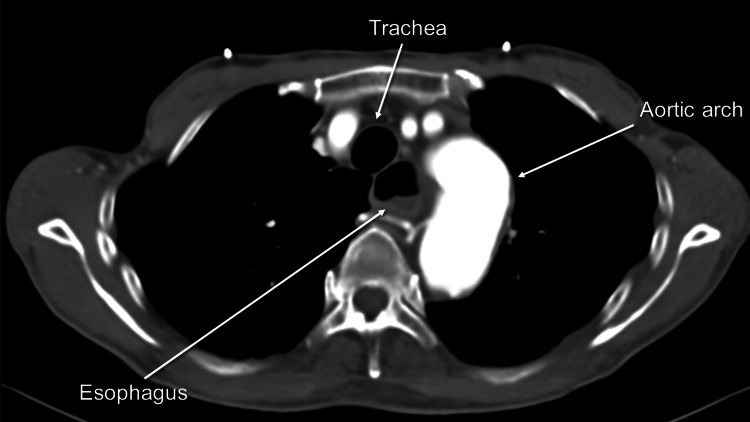
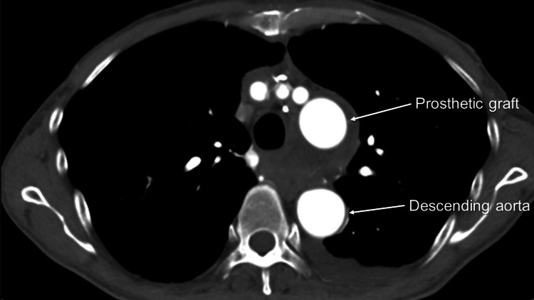
A 64-year-old man sought medical attention from a family physician, expressing concerns about dysphagia. Recognizing the complexity of the symptoms, the family physician promptly engaged the expertise of an attending physician at a regional hospital to ensure accurate diagnosis and management. Plain computed tomography (CT) revealed a space-occupied lesion located posterior to the trachea. Although mediastinal tumor was suspected at first, contrast-enhanced CT revealed a distal arch aneurysm that compressed the esophagus. The patient underwent total arch replacement, and the postoperative course was uneventful.

Keywords: bronchial artery; contrast-enhanced computed tomography; distal arch aneurysm; dysphagia; hoarseness; recurrent laryngeal nerve paralysis; vascular compression.


Conflict of interest statement

The authors have declared that no competing interests exist.

Figures

Figure 1
Figure 1. Plain computed tomography (CT) scan demonstrating a space occupying a lesion located posterior to the trachea
Figure 2
Figure 2. Contrast-enhanced computed tomography (CT) revealing dilated esophagus
Figure 3
Figure 3. Preoperative contrast-enhanced computed tomography (CT) revealing a distal arch aneurysm
Figure 4
Figure 4. Preoperative contrast-enhanced computed tomography (CT) showing bronchial arteries around a distal arch aneurysm
Figure 5
Figure 5. Preoperative contrast-enhanced computed tomography (CT)
Better visualized bronchial arteries in the coronal view
Figure 6
Figure 6. Postoperative contrast-enhanced computed tomography (CT) revealing a disappeared aneurysm

References

    1. Ascending thoracic aortic aneurysm elongation occurs in parallel with dilatation in a nonsurgical population. Gulati A, Zamirpour S, Leach J, et al. Eur J Cardiothorac Surg. 2023;63 - PMC - PubMed
    1. A patient with dysphagia due to an aortic aneurysm. Kim JH, Jang SW, Kim DB, et al. Korean Circ J. 2009;39:258–260. - PMC - PubMed
    1. Pictorial review of the pulmonary vasculature: from arteries to veins. Marini TJ, He K, Hobbs SK, Kaproth-Joslin K. Insights Imaging. 2018;9:971–987. - PMC - PubMed
    1. Hoarseness as the initial symptom of aortic arch aneurysm. Brunhuber C, Le Borgne P. Ann Thorac Surg. 2016;102:0. - PubMed
    1. Recurrent aspiration pneumonia due to bilateral associated laryngeal paralysis after tooth extraction under general anesthesia: a case report. Koshika K, Ouchi T, Serita R, Koitabashi T. JA Clin Rep. 2020;6:29. - PMC - PubMed

Publication types

LinkOut - more resources