Review
. 2023 Jul-Dec;27(9):793-806.
doi: 10.1080/14728222.2023.2259099. Epub 2023 Sep 14.

Fatty acid-mediated signaling as a target for developing type 1 diabetes therapies

Affiliations
Review

Fatty acid-mediated signaling as a target for developing type 1 diabetes therapies

Ivo Díaz Ludovico et al. Expert Opin Ther Targets. 2023 Jul-Dec.

Abstract

Introduction: Type 1 diabetes (T1D) is an autoimmune disease in which pro-inflammatory and cytotoxic signaling drive the death of the insulin-producing β cells. This complex signaling is regulated in part by fatty acids and their bioproducts, making them excellent therapeutic targets.

Areas covered: We provide an overview of the fatty acid actions on β cells by discussing how they can cause lipotoxicity or regulate inflammatory response during insulitis. We also discuss how diet can affect the availability of fatty acids and disease development. Finally, we discuss development avenues that need further exploration.

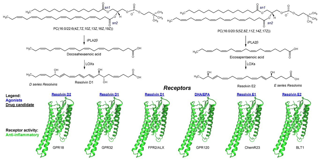
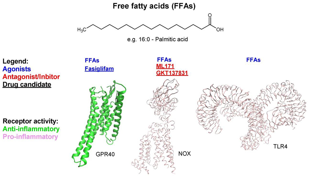
Expert opinion: Fatty acids, such as hydroxyl fatty acids, ω-3 fatty acids, and their downstream products, are druggable candidates that promote protective signaling. Inhibitors and antagonists of enzymes and receptors of arachidonic acid and free fatty acids, along with their derived metabolites, which cause pro-inflammatory and cytotoxic responses, have the potential to be developed as therapeutic targets also. Further, because diet is the main source of fatty acid intake in humans, balancing protective and pro-inflammatory/cytotoxic fatty acid levels through dietary therapy may have beneficial effects, delaying T1D progression. Therefore, therapeutic interventions targeting fatty acid signaling hold potential as avenues to treat T1D.

Keywords: Type 1 diabetes; cell signaling; lipid mediators; saturated fatty acids; therapeutic targets; β-cell death; β-cell protection; ω-3 fatty acids; ω-6 fatty acids.


Conflict of interest statement

The authors have no relevant affiliations or financial involvement with any organization or entity with a financial interest in or financial conflict with the subject matter or materials discussed in the manuscript. This includes employment, consultancies, honoraria, stock ownership or options, expert testimony, grants, or patents received or pending, or royalties.

Figures

Figure 1 –
Figure 1 –. Structure and nomenclature of fatty acids.
The figure shows examples of A) saturated, B) branched and, D) unsaturated fatty acids. An example of desaturation reaction is shown in D). Double bonds are named based on their position relative to the Δ carbon or classes of unsaturated fatty acids are named based on the position of the ω carbon. “E” or “Z” is designated to specify the trans and cis stereochemistry, respectively. Abbreviations: CoA, Coenzyme A; FAHFA, fatty acid esters of hydroxy fatty acid.
Figure 2.
Figure 2.. Arachidonic acid pathway.
The pathway shows examples of bioactive products of arachidonic acid oxidation along with their binding receptors. Receptors are colored based on their pro- or anti-inflammatory activity or both. Agonists (blue), inhibitors/antagonists (red), and drug candidates (underlined) are highlighted. Abbreviations: ALX, lipoxin A4 receptor; COX, cyclooxygenase; CYP, cytochrome P450; CysLT1, cysteinyl leukotriene receptor 1; CysLT2, cysteinyl leukotriene receptor 2; DHET, dihydroxyeicosatrienoic acid; DP, prostaglandin D2 receptor; EET, epoxyeicosatrienoic acid; EP1, prostaglandin E2 receptor 1; EP2, prostaglandin E2 receptor 2; EP3, prostaglandin E2 receptor 3; EP4, prostaglandin E2 receptor 4; FP, prostaglandin E2 receptor; FPR2, formyl peptide receptor 2; HETE, hydroxyeicosatetraenoic acid; IP, protacylcin receptor; iPLA2β; Calcium-independent phospholipase A2β; LT, leukotriene; LTB4R; leukotriene B4 receptor; LTB4R2, leukotriene B4 receptor 2; LOX, lipoxygenase; NOX, NADPH oxidase; PG, prostraglandin; TP, thromboxane A2 receptor.
Figure 3 –
Figure 3 –. ω-3 fatty acid pathway.
The pathway shows examples of bioactive lipids of ω-3 fatty acid pathway along with their binding receptors. Agonists (blue), and drug candidates (underlined) are highlighted. Receptors are colored based on their pro-inflammatory activity. Abbreviations: ALX, lipoxin A4 receptor; BLT1, leukotriene B4 receptor 1; ChemR23, Chemerin-like receptor 1; DHA, docosahexaenoic acid; EPA, eicosapentaenoic acid; FPR2, formyl peptide receptor 2; GPR18, G-protein coupled receptor 18; GPR32, G-protein coupled receptor 32; GPR120, G-protein coupled receptor 120; iPLA2β; Calcium-independent phospholipase A2β; LOX, lipoxygenase.
Figure 4 –
Figure 4 –. Free fatty acid pathway.
The pathway shows an example of bioactive lipid, palmitic acid, along with binding receptors and an enzyme. Receptors and enzymes are colored based on their pro-inflammatory activity. Agonists (blue), inhibitors (red), and drug candidates (underlined) are highlighted. Abbreviations: FFAs, free fatty acids; GPR40, G-protein coupled receptor 40; NOX, NADPH oxidase; TLR4, Toll-like receptor 4.
Figure 5 –
Figure 5 –. Fatty acid ester of hydroxy fatty acid (FAHFA) pathway.
The pathway shows the formation of FAHFAs along with their binding receptors. Agonists (blue), and drug candidates (underlined) are highlighted. Receptors are colored based on their pro-inflammatory activity. Abbreviations: FAHFA, fatty acid ester of hydroxy fatty acid; GPR40, G-protein coupled receptor 40; GPR120, G-protein coupled receptor 120.

References

    1. Gregory GA, Robinson TIG, Linklater SE, et al. Global incidence, prevalence, and mortality of type 1 diabetes in 2021 with projection to 2040: a modelling study. Lancet Diabetes Endocrinol. 2022. Oct;10(10):741–760. - PubMed
    1. DiMeglio LA, Evans-Molina C, Oram RA. Type 1 diabetes. Lancet. 2018. Jun 16;391(10138):2449–2462. - PMC - PubMed
    1. Eizirik DL, Colli ML, Ortis F. The role of inflammation in insulitis and beta-cell loss in type 1 diabetes. Nat Rev Endocrinol. 2009. Apr;5(4):219–26. - PubMed
    1. Lei X, Zhang S, Emani B, et al. A link between endoplasmic reticulum stress-induced beta-cell apoptosis and the group VIA Ca2+-independent phospholipase A2 (iPLA2beta). Diabetes Obes Metab. 2010. Oct;12 Suppl 2(0 2):93–8. - PMC - PubMed
    1. Nelson AJ, Stephenson DJ, Bone RN, et al. Lipid mediators and biomarkers associated with type 1 diabetes development. JCI Insight. 2020. Aug 20;5(16). - PMC - PubMed

Publication types

LinkOut - more resources