. 2023 Sep 1;64(12):27.
doi: 10.1167/iovs.64.12.27.

Regulation of Axon Guidance by Slit2 and Netrin-1 Signaling in the Lacrimal Gland of Aqp5 Knockout Mice

Affiliations

Regulation of Axon Guidance by Slit2 and Netrin-1 Signaling in the Lacrimal Gland of Aqp5 Knockout Mice

Ying Bai et al. Invest Ophthalmol Vis Sci. .

Abstract

Purpose: Dry eye disease (DED) is multifactorial and associated with nerve abnormalities. We explored an Aquaporin 5 (AQP5)-deficiency-induced JunB activation mechanism, which causes abnormal lacrimal gland (LG) nerve distribution through Slit2 upregulation and Netrin-1 repression.

Methods: Aqp5 knockout (Aqp5-/-) and wild-type (Aqp5+/+) mice were studied. LGs were permeabilized and stained with neuronal class III β-tubulin, tyrosine hydroxylase (TH), vasoactive intestinal peptide (VIP), and calcitonin gene-related peptide (CGRP). Whole-mount images were acquired through tissue clearing and 3D fluorescence imaging. Mouse primary trigeminal ganglion (TG) neurons were treated with LG extracts and Netrin-1/Slit2 neutralizing antibody. Transcription factor (TF) prediction and chromatin immunoprecipitation-polymerase chain reaction (ChIP-PCR) experiments verified the JunB binding and regulatory effect on Netrin-1 and Slit2.

Results: Three-dimensional tissue and section immunofluorescence showed reduced LG nerves in Aqp5-/- mice, with sympathetic and sensory nerves significantly decreased. Netrin-1 was reduced and Slit2 increased in Aqp5-/- mice LGs. Aqp5+/+ mice LG tissue extracts (TEs) promoted Aqp5-/- TG neurons axon growth, but Netrin-1 neutralizing antibody (NAb) could inhibit that promotion. Aqp5-/- mice LG TEs inhibited Aqp5+/+ TG axon growth, but Slit2 NAb alleviated that inhibition. Furthermore, JunB, a Netrin-1 and Slit2 TF, could bind them and regulate their expression. SR11302, meanwhile, reversed the Netrin-1 and Slit2 shifts caused by AQP5 deficiency.

Conclusions: AQP5 deficiency causes LG nerve abnormalities. Persistent JunB activation, the common denominator for Netrin-1 suppression and Slit2 induction, was found in Aqp5-/- mice LG epithelial cells. This affected sensory and sympathetic nerve fibers' distribution in LGs. Our findings provide insights into preventing, reversing, and treating DED.


Conflict of interest statement

Disclosure: Y. Bai, None; G. Di, None; H. Ge, None; B. Li, None; K. Zhang, None; D. Zhang, None; D. Wang, None; P. Chen, None

Figures

Figure 1.
Figure 1.
Abnormal LG nerves in Aqp5/− mice. (A) The unsectioned LGs of adult mice and Aqp5−/− mice were processed for tubulin βIII, TH, VIP, and CGRP immunolabeling. The 3D projection image of tissue at 2.5 times (left panel) and 10 times (right panel) magnification of the light sheet image are shown. Scale bars = 800 µm (2.5x) and 200 µm (10x). (B) The ratio of nerve volume to total volume of Aqp5+/+ and Aqp5−/− lacrimal gland was quantified analysis (n = 5; mean ± SD; n.s., not significant; *P < 0.05; **P < 0.01; Student's t-test).
Figure 2.
Figure 2.
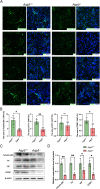
Lower sympathetic and sensory nerve densities in the LG of Aqp5/ mice. (A) Representative photomicrographs of the LGs from adult Aqp5+/+ and Aqp5−/− mice immunostained for tubulin βIII, TH, VIP, CGRP (green) and DAPI-stained cell nuclei (blue). Scale bars = 20 µm. (B) Positive analyses of immunofluorescence staining presented in (A). The significance of the difference between Aqp5+/+ and Aqp5−/− mice in respective nerve group is presented (n = 4; mean ± SD; n.s., not significant; *P < 0.05; **P < 0.01; Mann-Whitney test). (C) Western blot analysis of content of protein of tubulin βIII, TH, VIP, and CGRP in LG tissues lysates from Aqp5+/+ and Aqp5−/− adult mice. (D) Densitometric analyses of Western blots presented in (C). The significance of the difference between Aqp5+/+ and Aqp5−/− mice in respective nerve group is presented. Data are represented as means ± SD (n.s., not significant; *P < 0.05; **P < 0.01; Student's t-test).
Figure 3.
Figure 3.
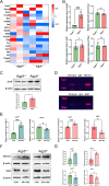
Decreased Netrin-1 and increased Slit2 in the LG of Aqp5/ mice. (A) Heatmap analysis of mRNA profiles about main neurotrophins and axon guidance cues in LG of Aqp5+/+ and Aqp5−/−. (B) The volcano plot for all expressed genes. Differentially expressed genes (P < 0.05) between Aqp5+/+ and Aqp5−/− LGs were presented with blue (down) and red (up). (C) Quantitative reverse transcription-polymerase chain reaction (qRT-PCR) analysis of the expression of Netrin-1 and Slit2 in LG tissues (n = 4; mean ± SD; *P < 0.05; *** P < 0.001). (D) Representative immunofluorescence images of LG tissue labeled with DAPI (blue), Netrin-1, and Slit2 (green). Scale bars = 20 µm. (E) Positive analyses of immunofluorescence staining presented in (D) (n = 6; mean ± SD; n.s., not significant; *P < 0.05; ***P < 0.001; Mann-Whitney test). (F) Western blot analysis of content of protein of Netrin-1 and Slit2 in LG tissues lysates from Aqp5+/+ and Aqp5−/− adult mice. (G) Densitometric analyses of Western blots presented in (F) (n = 3; mean ± SD; *P < 0.05; Student's t-test).
Figure 4.
Figure 4.
Netrin-1 reduction and Slit2 elevation in primary LG epithelial cells of Aqp5/ mice. (A) Aqp5+/+ and Aqp5−/− LG epithelial cells were fixed and immunolabeled with Netrin-1 (green), Slit2 (green), and DAPI (blue). Scale bars = 10 µm. (B) Positive analyses of immunofluorescence staining presented in (A) (n = 5; mean ± SD; ***P < 0.001; Student's t-test). (C) Western blot analysis of content of protein of Netrin-1 and Slit2 in LG epithelial cells from Aqp5+/+ and Aqp5−/− adult mice. (D) Densitometric analyses of Western blots presented in (C) (n = 3; mean ± SD; **P < 0.01; ***P < 0.001; Student's t-test).
Figure 5.
Figure 5.
Netrin-1 and Slit2 induced TG axonal extension and repulsion. (A) Axonal growth of Aqp5+/+ and Aqp5/ TG neurons after addition of rmNetrin-1 and rmSlit2, respectively. Scale bars = 25 µm. (B) Quantitative statistics of maximum branch length of (A) (n = 8; mean ± SD; *P < 0.05; **P < 0.01; Mann-Whitney test).
Figure 6.
Figure 6.
The effect of TEs of LGs on the outgrowth of TG axons. (A) Axonal growth of Aqp5+/+ TG neurons after addition of tissue extractions from Aqp5/ LGs and the Axon growth of Aqp5/ TG cells after adding tissue extractions from Aqp5+/+ LGs. (B) Quantitative statistics of maximum branch length of (A) (n = 8; mean ± SD; *P < 0.05; **P < 0.01; Student's t-test). (C) Axonal growth of Aqp5+/+ TG cells after addition of TEs from Aqp5/ LGs and Slit2 NAbs at the same time. (D) Quantitative statistics of maximum branch length of (C) (n = 8; mean ± SD; ***P < 0.001; Student's t-test). (E) Axonal growth of Aqp5/ TG cells after addition of TEs from Aqp5+/+ LGs and Netrin-1 NAbs at the same time. (F) Quantitative statistics of maximum branch length of (E) (n = 8; mean ± SD; **P < 0.01; Student's t-test). Scale bars = 25 µm.
Figure 7.
Figure 7.
JunB binds to and regulates Netrin-1 and Slit2 in Aqp5/ mice LG epithelial cells. (A) Heatmap analysis of mRNA profiles about transcription factors that co-transcribe Netrin-1 and Slit2 in LG of Aqp5+/+ and Aqp5−/−. (B) The qRT-PCR analysis of the expression of JunB, Jun, JunD, and Fosl in LG tissues (n = 4; mean ± SD; n.s., not significant; *** P < 0.001; Student's t-test). (C) Western blotting analysis showed the expression level JunB in LG epithelial cells. (D) ChIP-PCR analyses of JunB enrichment at binding sites in the murine Netrin-1 and Slit2 promoter. (E) The qRT-PCR analysis of the expression of Netrin-1 and Slit2 in Aqp5+/+ and Aqp5/ LG epithelial cells treated with SR11302 or DMSO 24 hours (n = 4; mean ± SD; * P < 0.05; *** P < 0.001; Student's t-test and Mann-Whitney test). (F) Western blot analysis of content of protein of Netrin-1 and Slit2 in Aqp5+/+ and Aqp5/ LG epithelial cells treated with SR11302 or DMSO 24 hours. (G) Densitometric analyses of Western blots presented in (F) (n = 3; mean ± SD; * P < 0.05; ** P < 0.01; Student's t-test)
Figure 8.
Figure 8.
Schema of proposed mechanisms of axonal changes in LGs induced by AQP5 deficiency.

Similar articles

Cited by

References

    1. Willcox MDP, Argüeso P, Georgiev GA, et al. .. TFOS DEWS II Tear Film Report. Ocul Surf . 2017; 15(3): 366–403. - PMC - PubMed
    1. Rolando M, Barabino S.. Dry eye disease: what is the role of vitamin D? Int J Mol Sci . 2023; 24(2): 1458. - PMC - PubMed
    1. Hou A, Bose T, Chandy KG, Tong L.. A chronic autoimmune dry eye rat model with increase in effector memory T cells in eyeball tissue. J Vis Exp . 2017;(124): 55592. - PMC - PubMed
    1. Pflugfelder SC, Stern ME.. Biological functions of tear film. Exp Eye Res . 2020; 197: 108115. - PMC - PubMed
    1. Stern ME, Beuerman RW, Fox RI, Gao J, Mircheff AK, Pflugfelder SC.. The pathology of dry eye: the interaction between the ocular surface and lacrimal glands. Cornea . 1998; 17(6): 584–589. - PubMed

Publication types