. 2023 Sep 26;42(9):113132.
doi: 10.1016/j.celrep.2023.113132. Epub 2023 Sep 13.

Multi-omics comparison of malignant and normal uveal melanocytes reveals molecular features of uveal melanoma

Affiliations

Multi-omics comparison of malignant and normal uveal melanocytes reveals molecular features of uveal melanoma

David Gentien et al. Cell Rep. .

Abstract

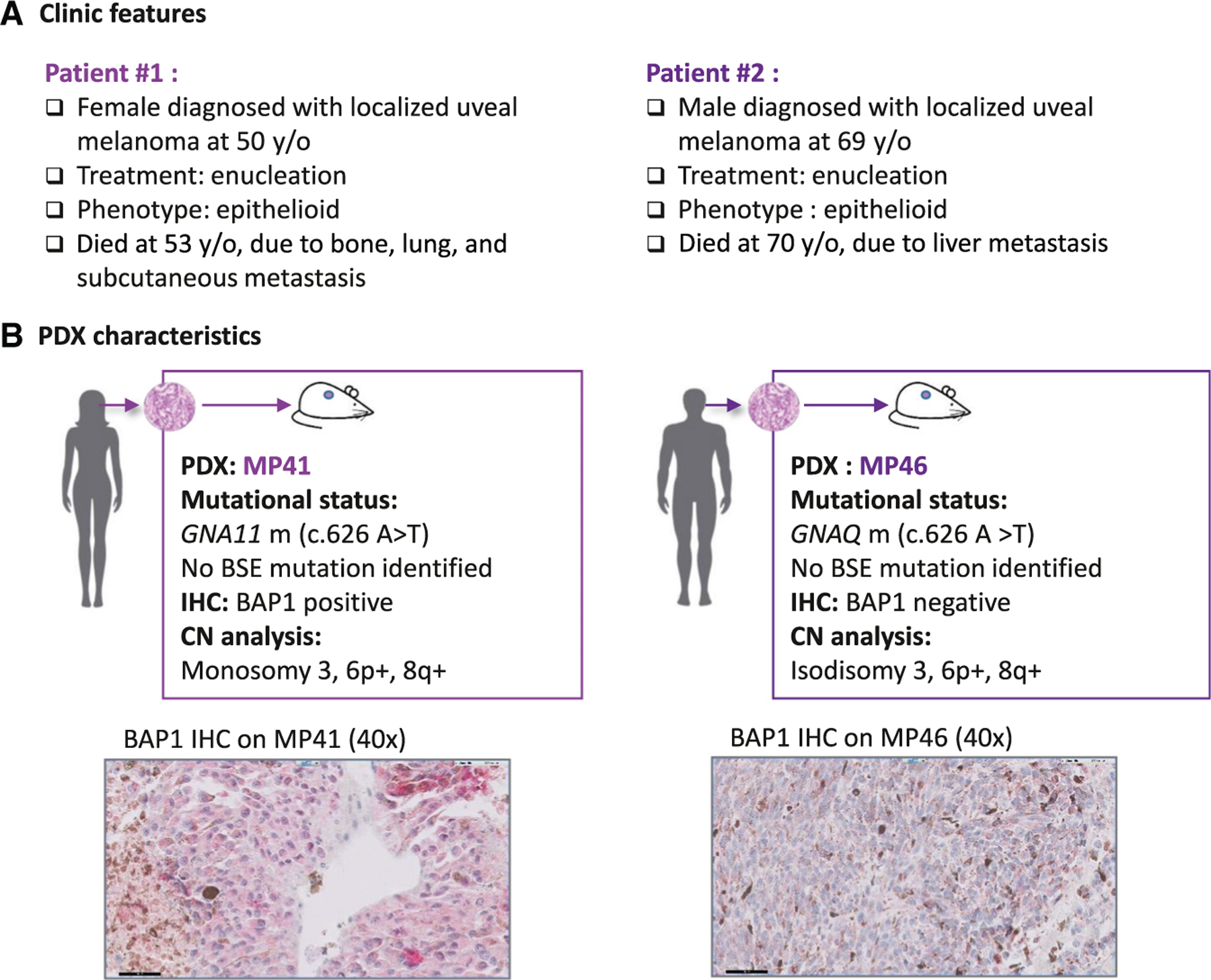
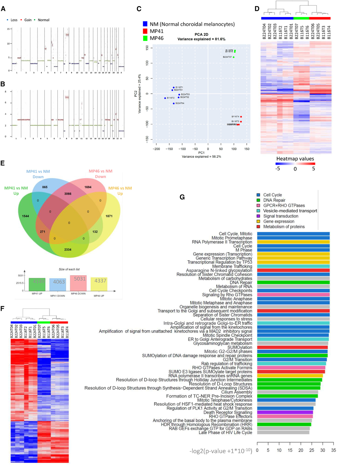
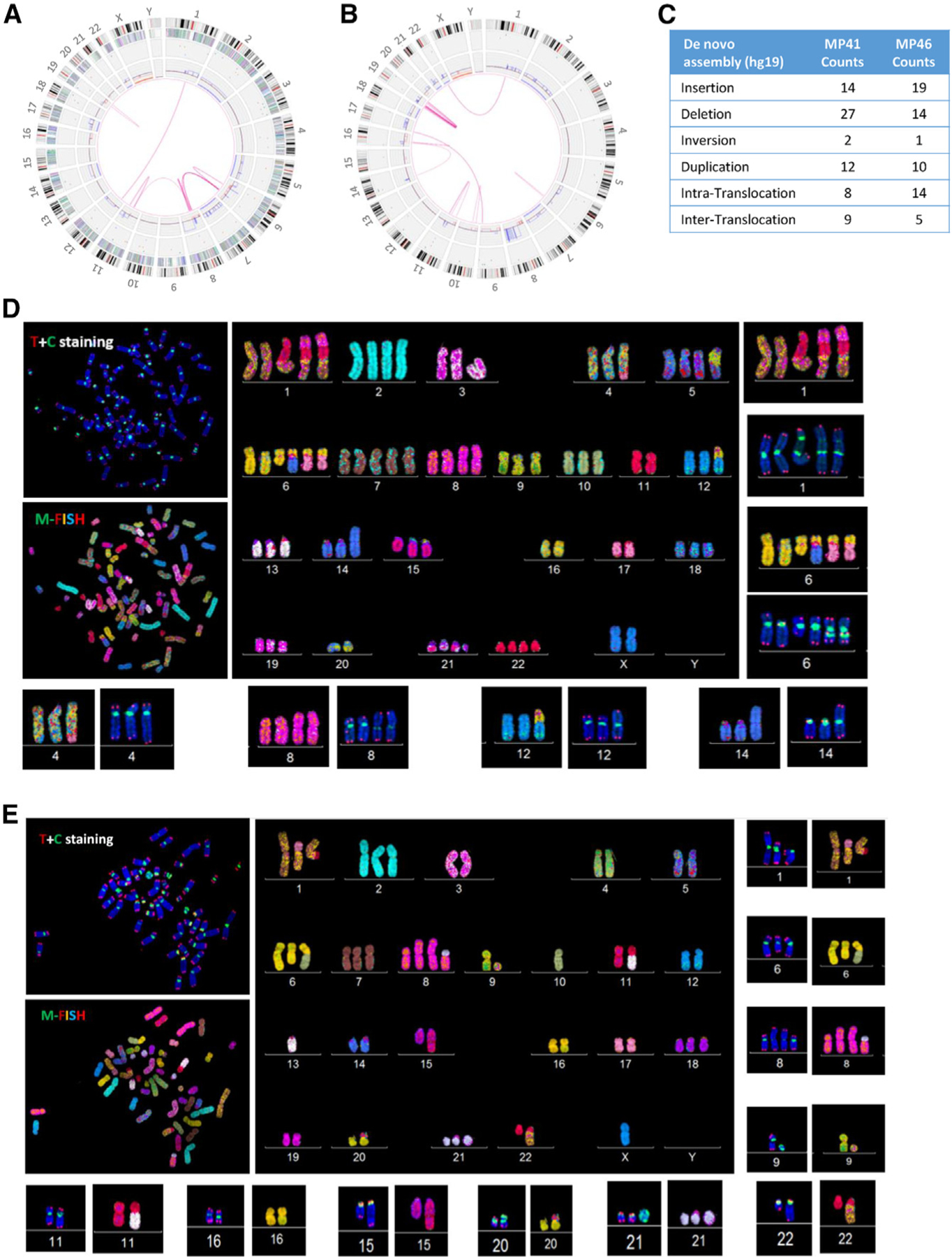
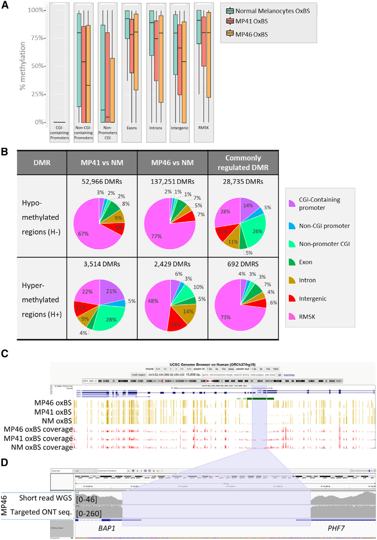
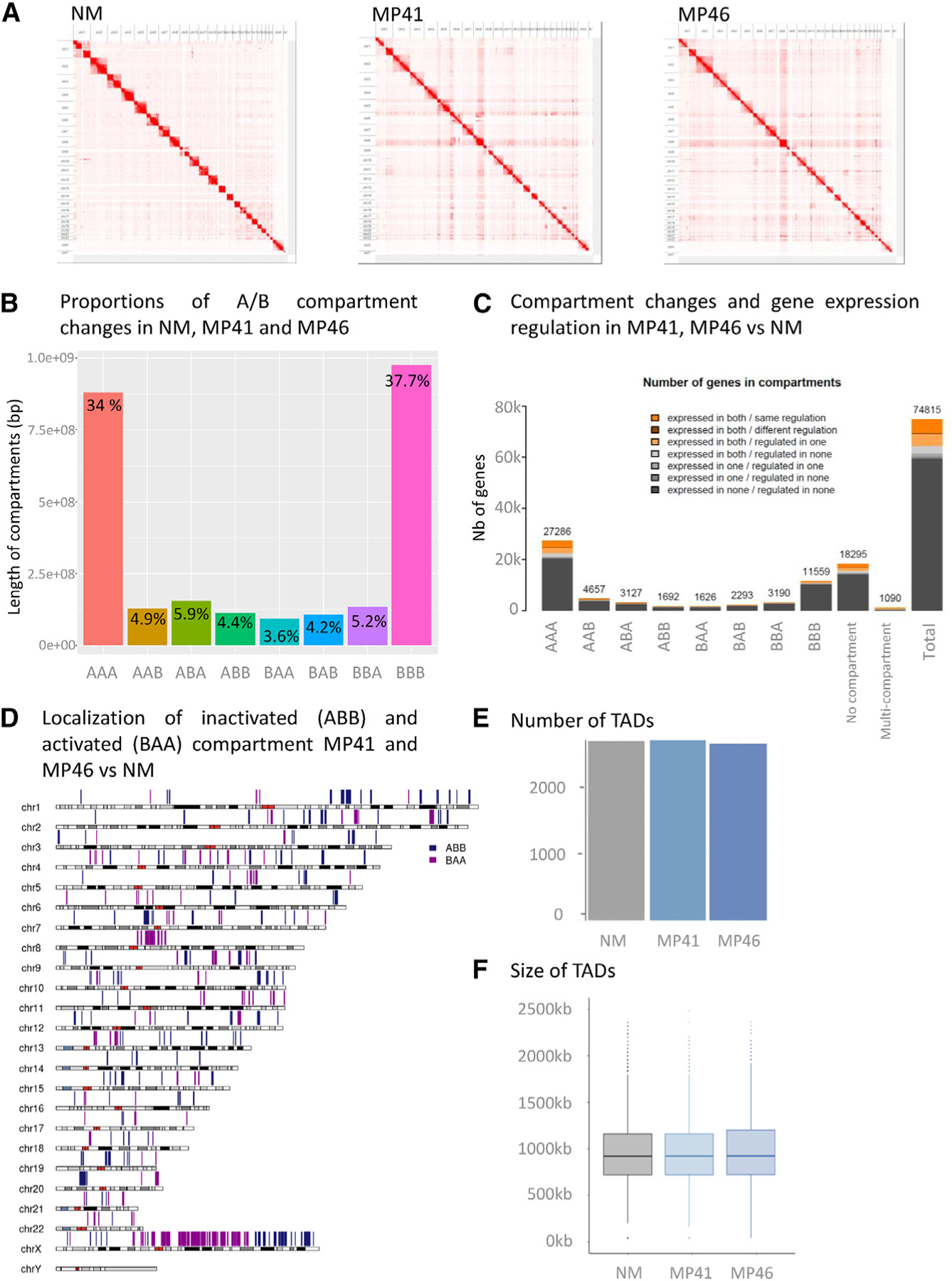
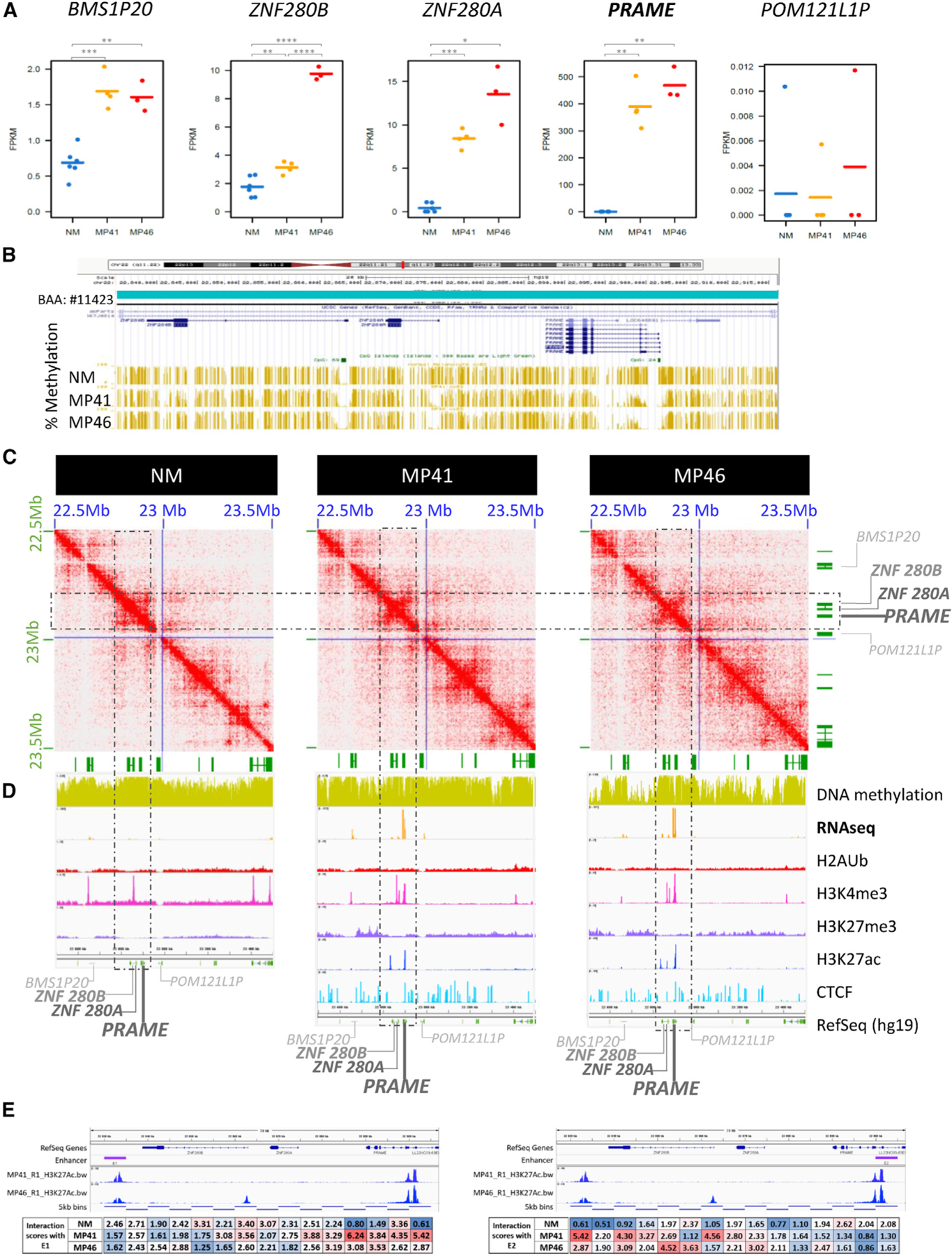
Uveal melanoma (UM) is a rare cancer resulting from the transformation of melanocytes in the uveal tract. Integrative analysis has identified four molecular and clinical subsets of UM. To improve our molecular understanding of UM, we performed extensive multi-omics characterization comparing two aggressive UM patient-derived xenograft models with normal choroidal melanocytes, including DNA optical mapping, specific histone modifications, and DNA topology analysis using Hi-C. Our gene expression and cytogenetic analyses suggest that genomic instability is a hallmark of UM. We also identified a recurrent deletion in the BAP1 promoter resulting in loss of expression and associated with high risk of metastases in UM patients. Hi-C revealed chromatin topology changes associated with the upregulation of PRAME, an independent prognostic biomarker in UM, and a potential therapeutic target. Our findings illustrate how multi-omics approaches can improve our understanding of tumorigenesis and reveal two distinct mechanisms of gene expression dysregulation in UM.

Keywords: BAP1; CP: Cancer; PRAME; genome instability; multi-omics; uveal melanoma.


Conflict of interest statement

Declaration of interests P.d.l.G. is a co-founder of Genosplice technology. A.J. and R.G. are employees of Genosplice technology. R.M. is the founder of Cell Environment.

Figures

Figure 1.
Figure 1.. Principal characteristics of MP41 and MP46 PDXs established from aggressive uveal melanomas
(A) Clinical characteristics of UM cases. (B) Main molecular characteristics of corresponding patient-derived xenograft models established and characterized previously. Mutational status was assessed with Sanger sequencing (GNAQ/GNA11, BAP1, SF3B1, EIF1AX), with Cytoscan HD microarrays for copy number analysis and BAP1 immunohistochemistry from formalin-fixed paraffin-embedded (FFPE) tissue section.
Figure 2.
Figure 2.. Genome and gene expression global overview
(A and B) MP41 (A) and MP46 (B) copy number profile established from whole-genome sequencing. Losses, gains, and normal regions are colored respectively in blue, red, and green. (C) Principal component analysis of RNA-seq of six normal choroidal melanocyte samples (blue), four preparations of MP41 UM cells (red). and three preparations of MP46 UM cells (green). (D) Hierarchical clustering of the same profiles. (E) Differential gene expression analysis of MP41 vs. NM and MP46 vs. NM to identify genes with a Log2 fold change greater than 1.5 and a p value lower than 0.05. (F) Heatmap of commonly regulated genes in MP41 vs. NM and MP46 vs. NM. (G) 50 most highly regulated pathways by reactome analysis of commonly regulated genes listed according to the significance (−log2 [p value + 1E-10]).
Figure 3.
Figure 3.. DNA optical mapping and FISH analysis of MP41 and MP46
(A and B) Circos plot of aberrations in (A) MP41 and (B) MP46. From the central to the periphery of the circos plot: whole-genome view summarizes intra- and inter-chromosomal translocations (pink lines), copy number gains and losses are listed on the first internal layer of the circos, and SVs (insertion, deletion inversion, and duplication) are labeled as colored dots in the intermediate layer of the circos. Gene density, cytobands, and chromosomes comprise the outer layers of the circos. (C) Number of insertions, deletions, inversions, and duplications and intra- and inter-translocations are detailed for MP41 and MP46 defined by Bionano optical mapping. (D and E) Telomere and M-FISH from analysis of (D) MP41 and (E) MP46 are derived from two different FISH analyses. Upper left panels show telomere (red signal) and centromere (green signal) staining and are counter labeled with DAPI (blue). Lower left panels show M-FISH analysis. Main panels correspond to the karyotype view.
Figure 4.
Figure 4.. Whole-genome DNA methylation analysis of UM models and normal melanocytes
(A) DNA methylation levels based on oxidative bisulfite DNA treatment followed by whole-genome sequencing are shown at CGI promoters, non-CGI promoters, non-promoters CGI, exons, introns, intergenic regions, and on repeats, in normal melanocytes, MP41, and MP46. (B) Differentially methylated regions (DMRs) as hypo- and hyper-DMRs (H− and H+) in 300-kb window in MP41 vs. NM and MP46 vs. NM. Commonly regulated DMRs correspond to DMRs identified in MP41 vs. NM and MP46 vs. NM comparisons. DMRs were considered as commonly regulated where sharing the same variation (H− or H+) and when their coordinates were identical or overlapping. (C) Percentage of CpG methylation in MP41, MP46, and NM in BAP1 locus through UCSC Genome Browser are represented in yellow, and sequencing coverages are represented in red bars. CpG island 129 overlaps the BAP1/PHF7 promoter. The blue area highlights the deletion detected in MP46. (D) IGV view of MP46 short-read sequencing (first line) and targeted ONT sequencing (second line) illustrate the boundaries of promoter/5′ UTR deletion in BAP1 and PHF7 genes.
Figure 5.
Figure 5.. Compartments and TADs in NMs and Ums
(A) Contacts maps derived from in situ Hi-C at the whole genome level for NM, MP41, and MP46. (B) Histogram of compartment changes in NM, MP41, and MP46. A and B compartments identified at 250-kb resolution. (C) Localization of inactivated (ABB) and activated (BAA) compartment in MP41 and MP46 vs. NM on a whole-genome view. (D) Integration of compartment changes and gene expression among NM, MP41, and MP46. (E and F) Number (E) and size (F) of TADs in NM, MP41, and MP46.
Figure 6.
Figure 6.. Multi-omics analysis of PRAME locus
(A) Gene expression of PRAME and its neighbors as BMS1P20, ZNF280B, and ZNF280A upstream genes and POM121LP downstream. RNA-seq data of NM, MP41, and MP46 replicates (FPKM). (B) UCSC Genome Browser view (hg19) of percentage of DNA methylation (golden bars). (C) DNA contacts maps of NM, MP41, and MP46 at 5-kb resolution in PRAME TAD (blue square). (D) UCSC Genome Browser view of the PRAME locus showing DNA methylation, RNA-seq (log2), H2AUb, H3K4me3, H3K27me3, H3K27Ac, CTCF, and RefSeq genes. (E) H3K27Ac marks and HiC interaction of PRAME promoter with potential distal enhancer E1. HiC interactions anchored in E1 (left) and E2 (right) quantified in boxes under genomic locus.

References

    1. Rodrigues M, Mobuchon L, Houy A, Alsafadi S, Baulande S, Mariani O, Marande B, Ait Rais K, Van der Kooij MK, Kapiteijn E, et al. (2019). Evolutionary Routes in Metastatic Uveal Melanomas Depend on MBD4 Alterations. Clin. Cancer Res 25, 5513–5524. 10.1158/1078-0432.CCR-19-1215. - DOI - PubMed
    1. Krantz BA, Dave N, Komatsubara KM, Marr BP, and Carvajal RD (2017). Uveal Melanoma: Epidemiology, Etiology, and Treatment of Primary Disease. Clin. Ophthalmol 11, 279–289. 10.2147/OPTH.S89591. - DOI - PMC - PubMed
    1. Van Raamsdonk CD, Bezrookove V, Green G, Bauer J, Gaugler L, O’Brien JM, Simpson EM, Barsh GS, and Bastian BC (2009). Frequent Somatic Mutations of GNAQ in Uveal Melanoma and Blue Naevi. Nature 457, 599–602. 10.1038/nature07586. - DOI - PMC - PubMed
    1. Van Raamsdonk CD, Griewank KG, Crosby MB, Garrido MC, Vemula S, Wiesner T, Obenauf AC, Wackernagel W, Green G, Bouvier N, et al. (2010). Mutations in GNA11 in Uveal Melanoma. N. Engl. J. Med 363, 2191–2199. 10.1056/NEJMoa1000584. - DOI - PMC - PubMed
    1. Moore AR, Ceraudo E, Sher JJ, Guan Y, Shoushtari AN, Chang MT, Zhang JQ, Walczak EG, Kazmi MA, Taylor BS, et al. (2016). Recurrent Activating Mutations of G-Protein-Coupled Receptor CYSLTR2 in Uveal Melanoma. Nat. Genet 48, 675–680. 10.1038/ng.3549. - DOI - PMC - PubMed

Publication types