

Microstructural white matter integrity in relation to vascular reactivity in Dutch-type hereditary cerebral amyloid angiopathy
- PMID: 37708241
- PMCID: PMC10925868
- DOI: 10.1177/0271678X231200425
Microstructural white matter integrity in relation to vascular reactivity in Dutch-type hereditary cerebral amyloid angiopathy
Abstract
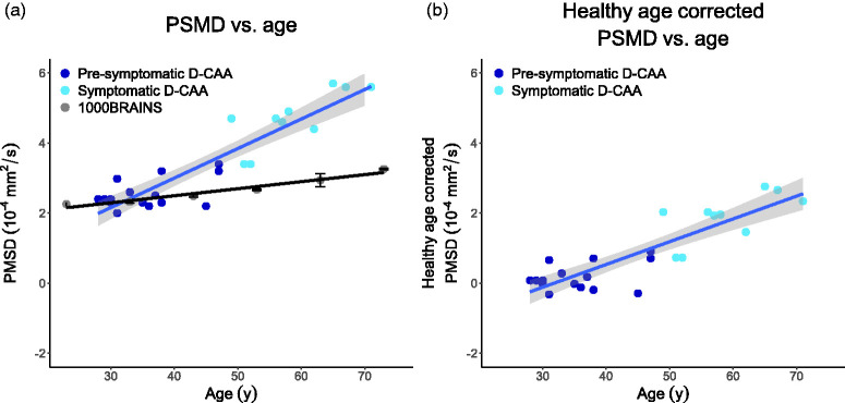
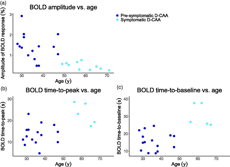
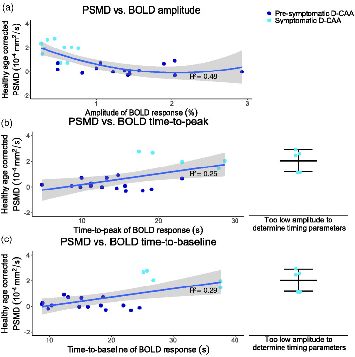
Cerebral Amyloid Angiopathy (CAA) is characterized by cerebrovascular amyloid-β accumulation leading to hallmark cortical MRI markers, such as vascular reactivity, but white matter is also affected. By studying the relationship in different disease stages of Dutch-type CAA (D-CAA), we tested the relation between vascular reactivity and microstructural white matter integrity loss. In a cross-sectional study in D-CAA, 3 T MRI was performed with Blood-Oxygen-Level-Dependent (BOLD) fMRI upon visual activation to assess vascular reactivity and diffusion tensor imaging to assess microstructural white matter integrity through Peak Width of Skeletonized Mean Diffusivity (PSMD). We assessed the relationship between BOLD parameters - amplitude, time-to-peak (TTP), and time-to-baseline (TTB) - and PSMD, with linear and quadratic regression modeling. In total, 25 participants were included (15/10 pre-symptomatic/symptomatic; mean age 36/59 y). A lowered BOLD amplitude (unstandardized β = 0.64, 95%CI [0.10, 1.18], p = 0.02, Adjusted R2 = 0.48), was quadratically associated with increased PSMD levels. A delayed BOLD response, with prolonged TTP (β = 8.34 × 10⁻⁶, 95%CI [1.84 × 10⁻⁶, 1.48 × 10⁻⁵], p = 0.02, Adj. R2 = 0.25) and TTB (β = 6.57 × 10⁻⁶, 95%CI [1.92 × 10⁻⁶, 1.12 × 10⁻⁵], p = 0.008, Adj. R2 = 0.29), was linearly associated with increased PSMD. In D-CAA subjects, predominantly in the symptomatic stage, impaired cerebrovascular reactivity is related to microstructural white matter integrity loss. Future longitudinal studies are needed to investigate whether this relation is causal.
Keywords: Diffusion magnetic resonance imaging; Dutch-type hereditary cerebral amyloid angiopathy; cerebral amyloid angiopathy; functional magnetic resonance imaging; vascular reactivity.
Conflict of interest statement
Declaration of conflicting interestsThe author(s) declared the following potential conflicts of interest with respect to the research, authorship, and/or publication of this article: M.R. Schipper reports independent support from the TRACK D-CAA consortium, consisting of Alnylam, Biogen, the Dutch CAA foundation, Vereniging HCHWA-D, and researchers from Leiden, Boston, and Perth.N. Vlegels reports support by the Dutch Heart Foundation.T.W. van Harten reports no disclosures.I. Rasing reports no disclosures.E.A. Koemans reports no disclosures.S. Voigt reports no disclosures.A. de Luca reports independent support from Alzheimer Nederland (WE-03-2022-11) as well as from ZonMW.K. Kaushik reports no disclosures.E.S. van Etten reports no disclosures.E.W. van Zwet reports no disclosures.G.M. Terwindt reports independent support from the Dutch Research Council (NWO), European Community, the Dutch Heart Foundation, the Dutch Brain Foundation, and the Dutch CAA foundation.G.J. Biessels reports support through the HBC (Heart-Brain Connection) Consortium, funded by the Netherlands CardioVascular Research Initiative, involving the Dutch Heart Foundation (CVON 2018-28 & 2012-06 Heart Brain Connection), Dutch Federation of University Medical Centers, the Netherlands Organization for Health Research and Development, and the Royal Netherlands Academy of Sciences.M.J.P. van Osch reports support by a NWO-VICI grant (016.160.351) and a NWO-Human Measurement Models 2.0 grant (18969) as well as support from the Dutch Research Council (NWO), European Community, the Dutch Heart Foundation, and the Dutch Brain Foundation.M.A.A. van Walderveen reports no disclosures.M.J.H. Wermer reports independent support from the Dutch Research Council (NWO) ZonMw (VIDI grant 91717337), the Netherlands Heart Foundation (Dekker grant 2016T86), and the Dutch CAA foundation.
Figures
References
-
- Vonsattel JP, Myers RH, Hedley-Whyte ETet al.. Cerebral amyloid angiopathy without and with cerebral hemorrhages: a comparative histological study. Ann Neurol 1991; 30: 637–649. - PubMed
-
- Charidimou A, Linn J, Vernooij MWet al.. Cortical superficial siderosis: detection and clinical significance in cerebral amyloid angiopathy and related conditions. Brain 2015; 138: 2126–2139. - PubMed
Publication types
MeSH terms
Supplementary concepts
LinkOut - more resources
Full Text Sources
