. 2023 Sep 15;381(6663):1176-1182.
doi: 10.1126/science.abp9556. Epub 2023 Sep 14.

MEG3 activates necroptosis in human neuron xenografts modeling Alzheimer's disease

Affiliations

MEG3 activates necroptosis in human neuron xenografts modeling Alzheimer's disease

Sriram Balusu et al. Science. .

Abstract

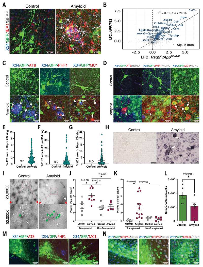
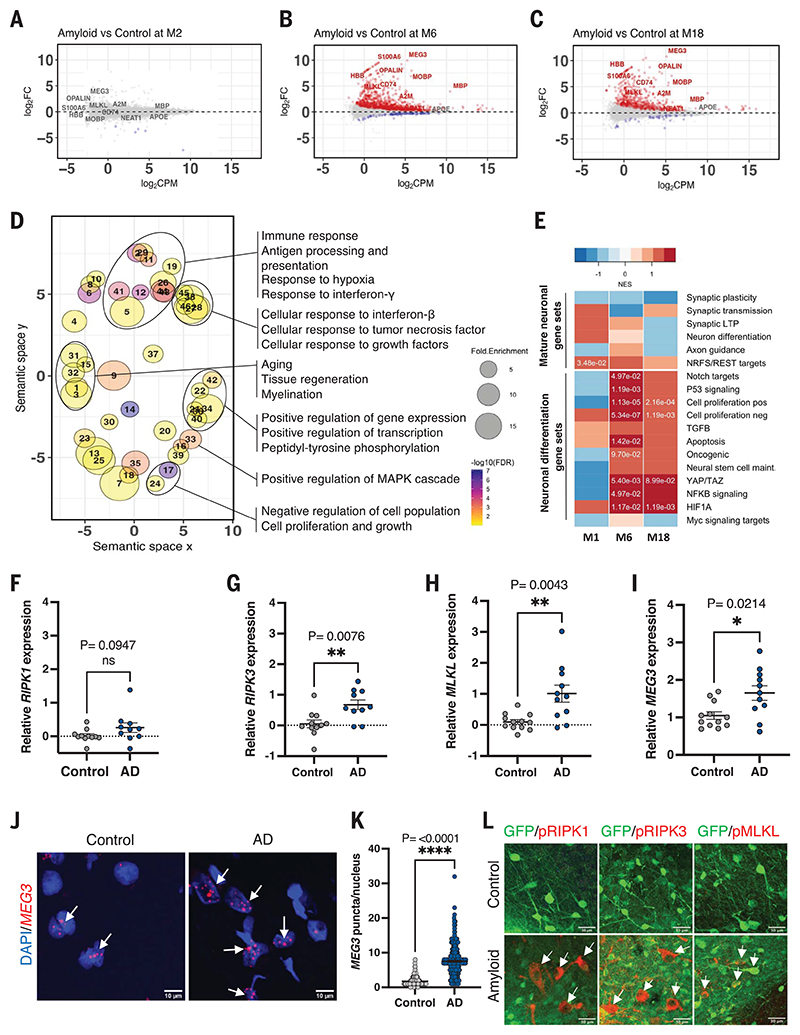
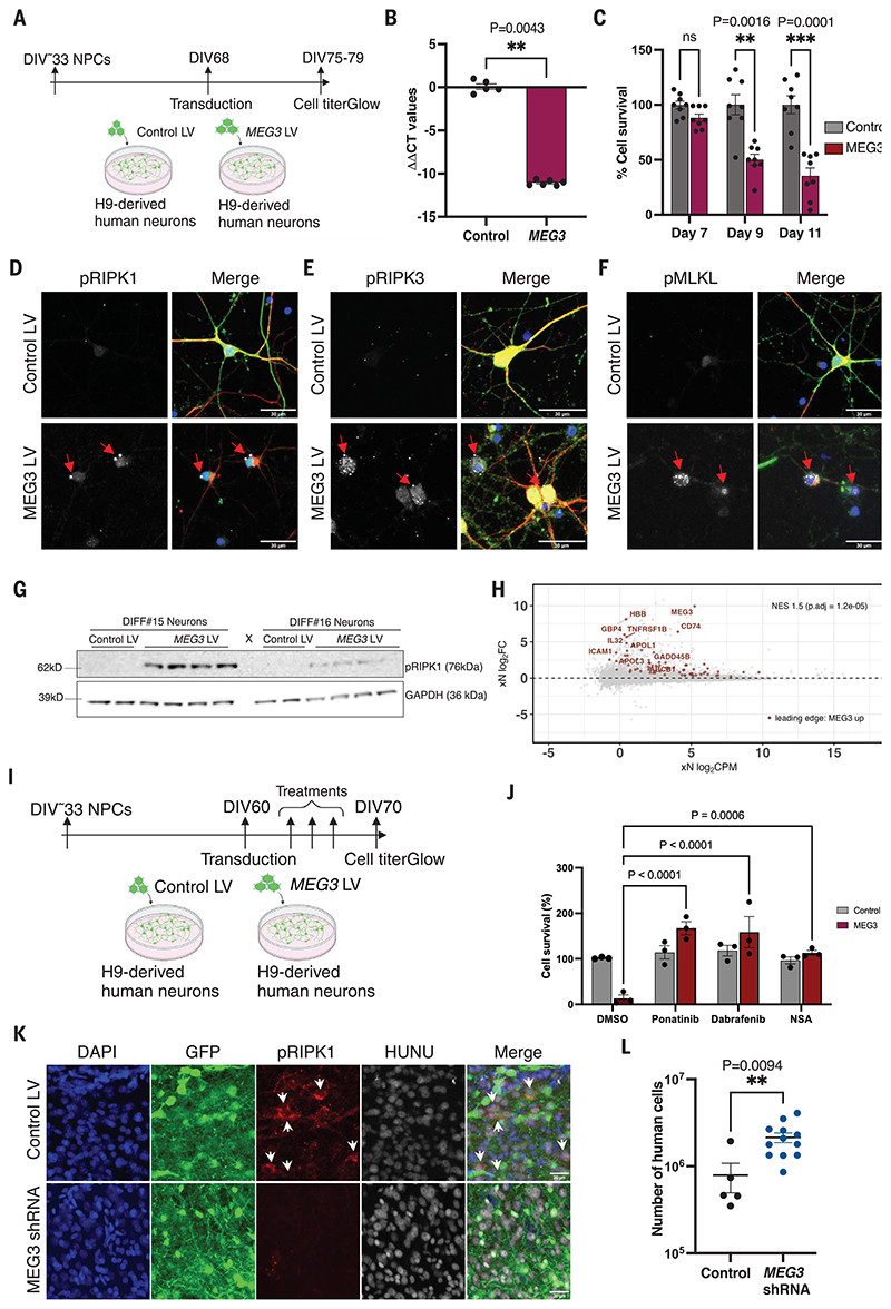
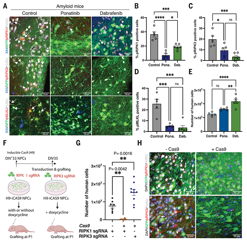
Neuronal cell loss is a defining feature of Alzheimer's disease (AD), but the underlying mechanisms remain unclear. We xenografted human or mouse neurons into the brain of a mouse model of AD. Only human neurons displayed tangles, Gallyas silver staining, granulovacuolar neurodegeneration (GVD), phosphorylated tau blood biomarkers, and considerable neuronal cell loss. The long noncoding RNA MEG3 was strongly up-regulated in human neurons. This neuron-specific long noncoding RNA is also up-regulated in AD patients. MEG3 expression alone was sufficient to induce necroptosis in human neurons in vitro. Down-regulation of MEG3 and inhibition of necroptosis using pharmacological or genetic manipulation of receptor-interacting protein kinase 1 (RIPK1), RIPK3, or mixed lineage kinase domain-like protein (MLKL) rescued neuronal cell loss in xenografted human neurons. This model suggests potential therapeutic approaches for AD and reveals a human-specific vulnerability to AD.


Conflict of interest statement

Competing interests: H.Z. has served on scientific advisory boards and/or as a consultant for AbbVie, Alector, Annexon, Artery Therapeutics, AZTherapies, Cognition Therapeutics, Denali, Eisai, NervGen, Pinteon Therapeutics, Red Abbey Labs, Passage Bio, Roche, Samumed, Siemens Healthineers, Triplet Therapeutics, and Wave; has given lectures in symposia sponsored by Cellectricon, Fujirebio, AlzeCure, Biogen, and Roche; and is a cofounder of Brain Biomarker Solutions in Gothenburg AB (BBS), which is a part of the GU Ventures Incubator Program. D.R.T. received speaker honoraria from Novartis Pharma Basel (Switzerland) and Biogen (USA); travel reimbursement from GE Healthcare (UK) and UCB (Belgium); and collaborated with GE Healthcare (UK), Novartis Pharma Basel (Switzerland), Probiodrug (Germany), and Janssen Pharmaceutical Company (Belgium). B.D.S. is or has been a consultant for Eli Lilly, Biogen, Janssen Pharmaceutical Company, Eisai, AbbVie, and other companies. B.D.S. is also a scientific founder of Augustine Therapeutics and a scientific founder and stockholder of Muna Therapeutics. W.-T.C. is an employee and stockholder of Muna Therapeutics.

Figures

Fig. 1
Fig. 1. Amyloid plaque deposition is sufficient to induce pathological tau in the grafts.
(A) Representative confocal images of grafted human neurons of 18-month-old control (n = 4) and amyloid (n = 4) mice. (B) Scatter plot comparing log fold changes (LFC) in APP/PS1 mice at 10 months and Rag2-/-/AppNL-G-F mice at 6 months old. Blue genes are significantly changed in both models (R2 = 0.81). (C) Representative confocal images showing NP-tau and neuronal soma tau (D) in 18-month-old control (n = 4) and amyloid (n = 4) mice. White arrows indicate X34 and NP-tau-positive cells. HUNU, human nucleus. Quantification of (E) AT8-positive tau, (F) PHF1-positive tau, (G) MC1-positive tau around Aβ plaque within 20 μm diameter in 18-month-old animals (n = 4, >100 plaques per mice). N.D, not detected. (H) Representative light microscope Gallyas silver stain images from 18-month-old control (n = 4) and amyloid (n = 4) grafted animals. (I) Electron micrograph image of immunogold-labeled sarkosyl insoluble tau fibrils isolated from 18-month-old control (n = 4) and amyloid (n = 4) mice. Tau fibrils are indicated with white (top panel) or green (bottom panel) arrows. (J) Quantification of plasma p-tau181 levels (every dot represents one mouse) and (K) p-tau231 levels from 18-month-old grafted and nongrafted mice. (L) Number of human neurons at 6 months after transplantation in control (n = 6) and amyloid (n = 6) mice using qPCR. (M) Confocal images taken from 6-month-old amyloid animals grafted with stem cell–derived mouse neurons (n = 4) did not stain with pathological tau markers (AT8, PHF1, or MC1) or (N) mouse-specific necrosome antibodies [pRIPK1(Ser166), pRIPK3 (Thr231/Thr232), or pMLKL (Ser345)]. The full panel, along with control mice, is shown in fig. S4, C and D. Scale bars: 30 μm [(A), (C), (D), (M), and (N)], 50 μm (H), 200 nm [(I), top row], 100 nm [(I), bottom row]. Values are presented as mean ± SEM. One-way analysis of variance (ANOVA) with Tukey’s post hoc test for multiple comparisons was used in (J) to (K), and Student’s t test was used in (M).
Fig. 2
Fig. 2. Transcriptional changes in xenografted neurons.
Bland-Altman MA plot showing differential expression of genes from RNA sequencing of human grafts from control and amyloid mice at (A) 2 months (control n = 5, amyloid n = 7), (B) 6 months (control n = 5, amyloid n = 5), and (C) 18 months (control n = 4, amyloid n = 3) after transplantation. Red indicates significantly up-regulated genes. Blue indicates significantly down-regulated genes [false discovery rate (FDR) < 0.05]. FC, fold change; CPM, counts per million. (D) GO analysis showing terms associated with genes up-regulated at 6 months. Point size represents the fold enrichment of up-regulated genes in the term, and color represents the -log10FDR. Only terms with FDR < 0.1 are shown. The x and y axes represent the “semantic space” (54). (E) Heatmap of normalized enrichment scores (NES) in neuronal dedifferentiation gene sets ranked along the differentially expressed genes in the xenografts (amyloid versus control) shown in (A) to (C). Positive enrichments in red, negative enrichments in blue. Significant FDR values (Padj < 0.01) are shown as numbers. (F to H) Analysis of (F) RIPK1, (G) RIPK3, and (H) MLKL mRNA expression using quantitative reverse transcription polymerase chain reaction (qRT-PCR) on AD (n = 11) and control (n = 10) postmortem human brain samples. (I) Analysis of MEG3 gene expression using qRT-PCR on AD (n = 11) and control (n = 10) postmortem human brain samples. (J) Confocal images showing MEG3 RNAscope in the temporal gyrus of AD (n = 3) and control (n = 2) postmortem human brain samples. Scale bars: 10 μm. (K) Number of MEG3 puncta per nucleus in control (>100 nuclei per sample, n = 2) and AD (>100 nuclei per sample, n = 3). (L) Representative confocal images showing the expression of activated necroptosis pathway markers pRIPK1, pRIPK3, or pMLKL in red (white arrows) in 18-month-old human neurons in control (n = 4) and amyloid (n = 4) mice. Scale bars: 30 μm. Values are presented as mean ± SEM. Student’s t test used in (F) to (I) and (K).
Fig. 3
Fig. 3. Long noncoding RNA MEG3 induces necroptosis in human neurons.
(A) Schematic representation of the MEG3 expression strategy using lentiviral vectors (LV) in H9-derived human neurons. (B) Analysis of the MEG3 expression 7 days after transduction with control LV (n = 5) or MEG3 LV (n = 6) in DIV75 neurons. (C) Analysis of neuronal cell survival using Cell-Titer-Glo reagent after transducing with either control LV (n = 8) or MEG3 LV (n = 8). (D) Confocal images showing pRIPK1 (Ser166), (E) pRIPK3 (Ser227), (F) pMLKL (Ser358) in neurons at DIV75 and 7 days after transduction with control LV (n = 3) or MEG3 LV (n = 3). Scale bars: 30 μm. (G) Immunoblot analysis of pRIPK1 (Ser166) levels 7 days after transduction (DIV75) with control LV (n = 4) or MEG3 LV (n = 8). Glyceraldehyde phosphate dehydrogenase (GAPDH) is the loading control. Two independent differentiations were analyzed. (H) Same Bland-Altman MA plot showing differential expression of bulk RNA sequencing of human grafts from 6 months, as in Fig. 2B. Genes highlighted in red are the leading-edge genes identified with GSEA, taking the top 400 up-regulated genes from the bulk sequencing of the primary neurons expressing MEG3 (fig. S10) and plotting them against the amyloid versus control fold changes of the human grafts (FDR < 0.05). (I) Schematic representation of the necroptosis inhibition in vitro using H9-derived human neurons (DIV60) with ponatinib (0.5 μM), dabrafenib (0.9 μM), and NSA (0.5 μM). (J) Analysis of neuronal cell survival using CellTiter-Glo reagent after necroptosis inhibitor treatment (n = 3). (K) Representative confocal images of grafted neurons showing pRIPK1 levels in samples obtained from the 6-month-old animals transplanted with control LV (n = 5) or the MEG3 shRNA (n = 5) transduced human NPCs. White arrows indicate pRIPK1-positive cells. Scale bars: 30 μm. (L) The human cell number was estimated from 6-month-old xenografted mice from control (n = 5) and MEG3 shRNA (n = 12) using qPCR. Values are presented as mean ± SEM. Student’s t test used in (B) and (L), one-way ANOVA with Tukey’s post hoc test for multiple comparisons used in (C), and two-way ANOVA with Dunnett’s multiple comparison test used in (J) to measure the statistical significance.
Fig. 4
Fig. 4. Inhibition of necroptosis prevents human neuronal cell loss in vivo.
(A) Confocal images showing activated necroptotic markers pRIPKl, pRIPK3, and pMLKL in amyloid animals (n = 5) and amyloid animals treated with ponatinib (n = 5) or dabrafenib (n = 6). White arrows indicate necroptosis marker-positive neurons. Scale bars: 30 μm. (B) Quantitative representation of percent of human cells immunoreactive to pRIPKl in control (n = 5) and ponatinib-treated (Pona.; n = 5) or dabrafenib-treated (Dab.; n = 6) animals. Control versus ponatinib: P ≤ 0.0001; control versus dabrafenib: P = 0.0007; ponatinib versus dabrafenib: P = 0.015. (C) Percent of human cells showing immunoreactivity to pRIPK3 in control (n = 5) and ponatinib-treated (n = 5) or dabrafenib-treated (n = 6) animals. Control versus ponatinib: P = 0.02; control versus dabrafenib: P = 0.0007; ponatinib versus dabrafenib: P = 0.2. (D) Quantitative representation of the percent of human cells showing immunoreactivity toward pMLKL in control (n = 5) and ponatinib-treated (n = 5) or dabrafenib-treated (n = 6) animals. Control versus ponatinib: P = 0.0003; control versus dabrafenib: P = 0.0001; ponatinib versus dabrafenib: P = 0.9. (E) Quantitative estimation of the number of human cells in control (n = 5) and ponatinib-treated (n = 5) or dabrafenib-treated (n = 6) grafted mice at 6 months after transplantation. Control versus ponatinib: not significant; control versus dabrafenib: P = 0.0001; ponatinib versus dabrafenib: P = 0.007. (F) Schematic representation of the RIPK1 and RIPK3 KO generation and transplantation. (G) Number of neurons surviving from 6-month-old mice transplanted with RIPK1 single guide RNA (sgRNA) without doxycycline (n = 7) or RIPK1 sgRNA with doxycycline (n = 7) or RIPK3 sgRNA with doxycycline (n = 10) using qPCR. (H) Representative immunofluorescence analysis of phosphorylated necroptosis markers pRIPK1 (n = 3) pRIPK3 (n = 3) from 6-month-old mice transplanted with RIPK1 sgRNA or RIPK3 sgRNA without and with Cas9 induction, as indicated. White arrows indicate pRIPK1- or pRIPK3-positive cells. Scale bars: 30 μm. Values are presented as mean ± SEM. One-way ANOVA with Tukey’s post hoc test for multiple comparisons was used in (B) to (E) and (G).

References

    1. He Z, et al. Nat Med. 2018;24:29–38. - PMC - PubMed
    1. Grueninger F, et al. Neurobiol Dis. 2010;37:294–306. - PubMed
    1. Mattsson-Carlgren N, et al. Sci Adv. 2020;6:eaaz2387. - PMC - PubMed
    1. Guela C, et al. Nat Med. 1998;4:827–831. - PubMed
    1. Choi SH, et al. Nature. 2014;515:274–278. - PMC - PubMed