Review
. 2023 Oct 10;56(10):2218-2230.
doi: 10.1016/j.immuni.2023.08.014. Epub 2023 Sep 13.

Dendritic cells as shepherds of T cell immunity in cancer

Affiliations
Review

Dendritic cells as shepherds of T cell immunity in cancer

Mikael J Pittet et al. Immunity. .

Abstract

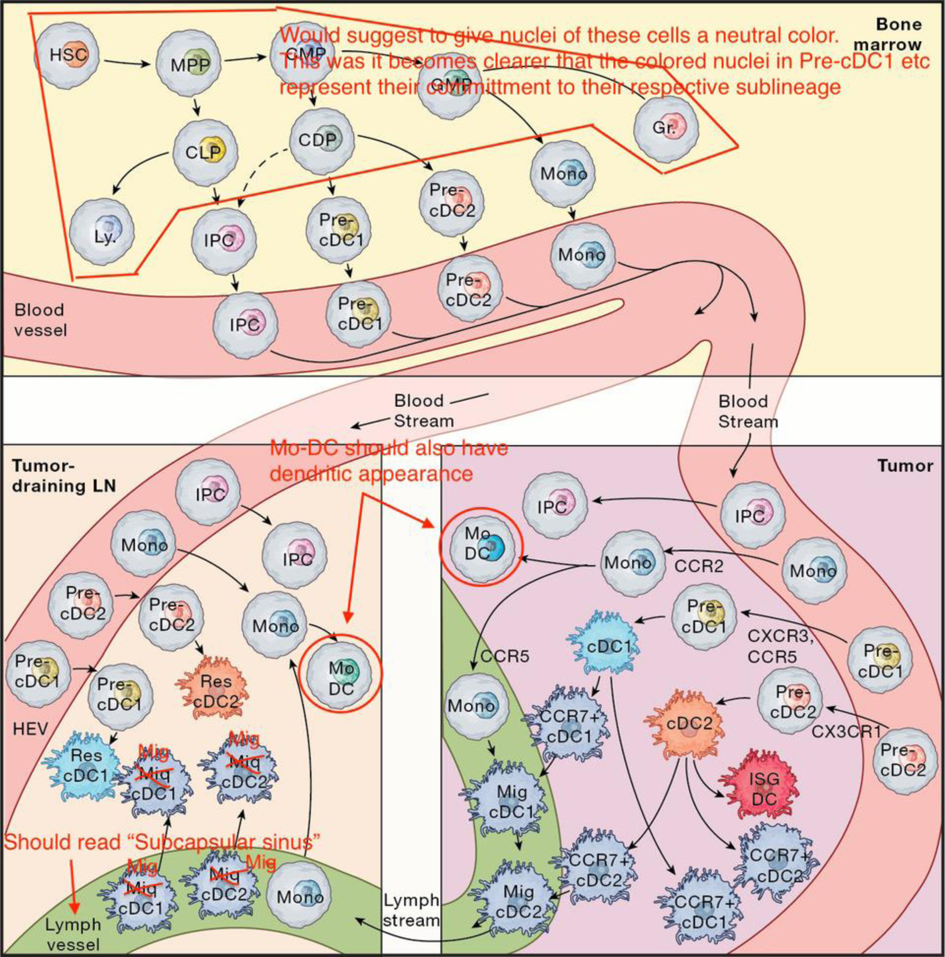
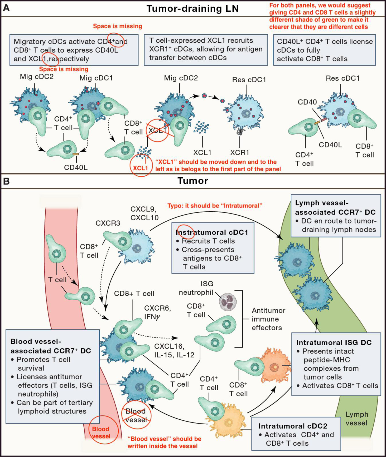
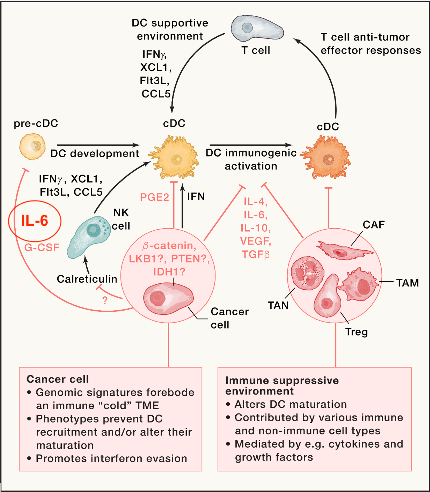
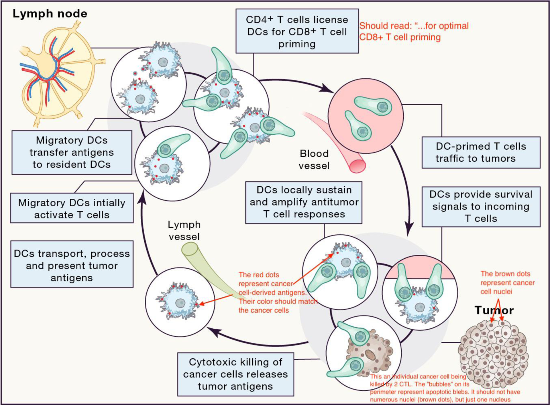
In cancer patients, dendritic cells (DCs) in tumor-draining lymph nodes can present antigens to naive T cells in ways that break immunological tolerance. The clonally expanded progeny of primed T cells are further regulated by DCs at tumor sites. Intratumoral DCs can both provide survival signals to and drive effector differentiation of incoming T cells, thereby locally enhancing antitumor immunity; however, the paucity of intratumoral DCs or their expression of immunoregulatory molecules often limits antitumor T cell responses. Here, we review the current understanding of DC-T cell interactions at both priming and effector sites of immune responses. We place emerging insights into DC functions in tumor immunity in the context of DC development, ontogeny, and functions in other settings and propose that DCs control at least two T cell-associated checkpoints of the cancer immunity cycle. Our understanding of both checkpoints has implications for the development of new approaches to cancer immunotherapy.


Conflict of interest statement

Declaration of interests M.J.P. has served as a consultant for Acthera, AstraZeneca, Debiopharm, Elstar Therapeutics, ImmuneOncia, KSQ Therapeutics, MaxiVax, Merck, Molecular Partners, Siamab Therapeutics, Third Rock Ventures, and Tidal. T.R.M. is a co-founder and consultant, and M.D.P. is a consultant for Monopteros Therapeutics. C.G. has served as a consultant for Cellino Biotech.

Figures

Figure 1
Figure 1. Cellular origins of tumor and lymph node DCs.
DCs are derived from myeloid progenitor cells in the bone marrow that traffic through blood to tumors and lymph nodes. In tumors they can adopt activated cell states such as CCR7+ DCs or interferon stimulated gene (ISG) DCs. CCR7+ DCs can either cluster around blood vessels to interact with blood extravasating T-cells, or move to lymphatics and become migratory DCs that traffic antigens to draining lymph nodes. Upon entering lymph nodes, migratory DCs initiate T-cell responses by priming naïve T-cells; setting off a complex interplay of antigen handoff to lymph node-resident DCs, and coordination of both cDC1 and cDC2 responses for successful antitumor immunity. HSC: hematopoietic stem cell; MPP: multipotent progenitor; CLP: common lymphoid progenitor; Ly: lymphocyte; IPC: interferon-producing cell; CMP: common myeloid progenitor; GMP: granulocyte monocyte progenitor; Gr: granulocyte; CDP: common dendritic progenitor; Mono: monocyte; MoDC: monocyte derived DC; migDC: migratory DC; resDC: resident DC; HEV: high endothelial venule.
Figure 2.
Figure 2.. Licensing of T-cell immunity by DCs in draining lymph nodes and tumors.
(A) Lymph Node: Migratory DCs interact with T-cells upon entering the lymph node from tumor-draining lymphatics. These interactions coordinate CD4+ and CD8+ T-cell responses and provide the basis for CD4+ T-cell “help”, such as through CD40L:CD40 stimulation, for CD8+ T-cell immune responses. T-cell-derived XCL1 can trigger recruitment of lymph node-resident cDC1s, enabling antigen transfer between migratory and resident DCs. CD4+ T-cell-“helped” DCs are fully licensed to robustly activate CD8+ T-cells. (B) Tumor: Intratumoral cDC1s (light blue) and cDC2s (orange) can profoundly influence the fate of incoming CD8+ and CD4+ T-cells and have distinct fates. For instance, some activated cDC1s and cDC2s can migrate via lymphatic vessels to tumor-draining lymph nodes, where they also contribute to T-cell responses. Other activated cDC1s and cDC2s remain in the tumor, where they can for example accumulate in perivascular niches near post-capillary venules of the tumor stroma and provide effector and survival signals to incoming T-cells. Some activated cDC2s can also acquire an interferon-stimulated gene (ISG) phenotype capable of activating CD8+ T-cells. The inserts illustrate some of the main functions ascribed to intratumoral cDCs. Key T-cell-derived immune signals (CXCR6, CXCR3, IFN-g), respond to and potentiate DC-derived signals (CXCL16, CXCL9, CXCL10, IL-12, and IL-15), resulting in the mutualistic support of T:DC interactions and sustained antitumor responses by multiple effector cells.
Figure 3.
Figure 3.. Suppression of DC immunogenic maturation by tumors.
DC development within tumors requires a supportive environment, which is driven at least in part by Flt3L, XCL1, CCL5, and IFN-g, which can be produced by various cell types, including natural killer (NK) cells. The latter can be activated by calreticulin following immunogenic tumor cell death. The release of type I interferons can also trigger DC-supportive environments. However, tumors can suppress these environments, for example, by suppressing NK cell activation or type I interferon production, or by releasing factors such as prostaglandin E2 (PGE2), interleukin-6, or granulocyte-colony stimulating factor (G-CSF) that block DC progenitor development. Tumor-derived factors can also condition an immunosuppressive microenvironment consisting of T regulatory (Treg) cells, cancer-associated fibroblasts (CAFs), tumor-associated macrophages (TAMs), and tumor-associated neutrophils (TANs). Together, these mechanisms block T:DC mutualistic interactions and promote evasion of antitumor immunity.
Figure 4.
Figure 4.. DCs participate in multiple T-cell-associated checkpoints in the cancer immunity cycle.
The original version of the cancer immunity cycle, which indicated the self-propagating generation of cancer immunity, was divided into several steps, starting with the release of tumor antigens from dying cancer cells and ending with the elimination of cancer cells by antitumor immune cells. Here, we further indicate that DCs participate in T-cell-associated checkpoints in ‘sub-circuits’ throughout the cancer immunity cycle, i.e. not only in tumor-draining lymph nodes where they prime naive antitumor T cells, but also in tumors where they drive effector differentiation and survival of incoming T cells. All these processes associated with DCs are necessary for successful antitumor immunity. Exploiting these checkpoints could be critical in the development of more effective cancer immunotherapies. Blue: DCs; green: T cells; light pink: tumor cells; red dots: tumor antigens.

References

    1. Schumacher TN, and Schreiber RD (2015). Neoantigens in cancer immunotherapy. Science 348, 69–74. 10.1126/science.aaa4971. - DOI - PubMed
    1. Finn OJ (2017). Human Tumor Antigens Yesterday, Today, and Tomorrow. Cancer Immunol Res 5, 347–354. 10.1158/2326-6066.CIR-17-0112. - DOI - PMC - PubMed
    1. Rizvi NA, Hellmann MD, Snyder A, Kvistborg P, Makarov V, Havel JJ, Lee W, Yuan J, Wong P, Ho TS, et al. (2015). Cancer immunology. Mutational landscape determines sensitivity to PD-1 blockade in non-small cell lung cancer. Science 348, 124–128. 10.1126/science.aaa1348. - DOI - PMC - PubMed
    1. Samstein RM, Lee CH, Shoushtari AN, Hellmann MD, Shen R, Janjigian YY, Barron DA, Zehir A, Jordan EJ, Omuro A, et al. (2019). Tumor mutational load predicts survival after immunotherapy across multiple cancer types. Nat Genet 51, 202–206. 10.1038/s41588-018-0312-8. - DOI - PMC - PubMed
    1. Spranger S, Luke JJ, Bao R, Zha Y, Hernandez KM, Li Y, Gajewski AP, Andrade J, and Gajewski TF (2016). Density of immunogenic antigens does not explain the presence or absence of the T-cell-inflamed tumor microenvironment in melanoma. Proc Natl Acad Sci U S A 113, E7759–E7768. 10.1073/pnas.1609376113. - DOI - PMC - PubMed

Publication types