Review
. 2023 Sep 15;8(1):352.
doi: 10.1038/s41392-023-01570-w.

Endoplasmic reticulum stress: molecular mechanism and therapeutic targets

Affiliations
Review

Endoplasmic reticulum stress: molecular mechanism and therapeutic targets

Xingyi Chen et al. Signal Transduct Target Ther. .

Abstract

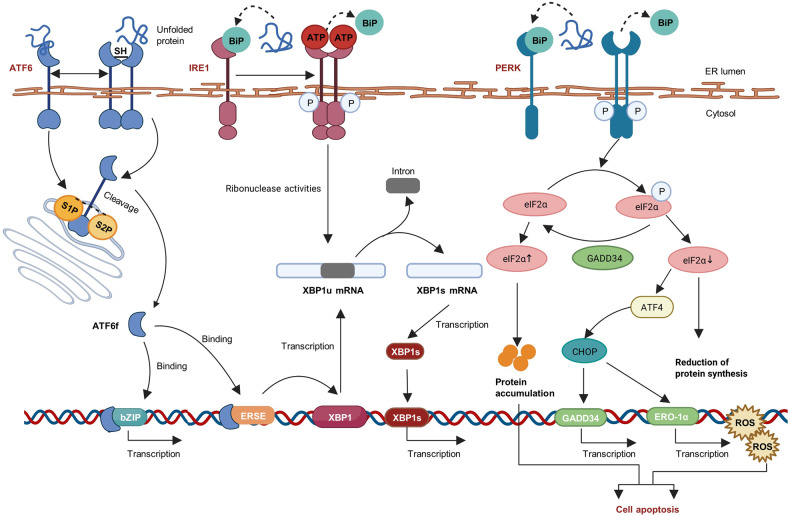
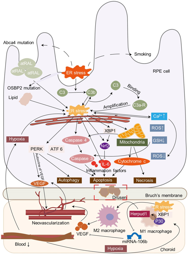
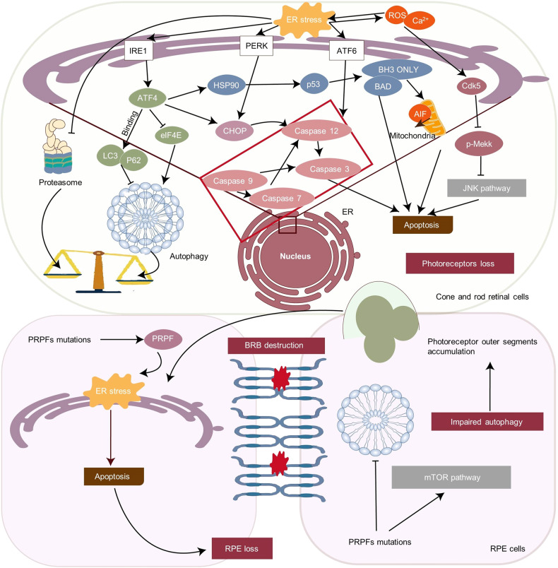
The endoplasmic reticulum (ER) functions as a quality-control organelle for protein homeostasis, or "proteostasis". The protein quality control systems involve ER-associated degradation, protein chaperons, and autophagy. ER stress is activated when proteostasis is broken with an accumulation of misfolded and unfolded proteins in the ER. ER stress activates an adaptive unfolded protein response to restore proteostasis by initiating protein kinase R-like ER kinase, activating transcription factor 6, and inositol requiring enzyme 1. ER stress is multifaceted, and acts on aspects at the epigenetic level, including transcription and protein processing. Accumulated data indicates its key role in protein homeostasis and other diverse functions involved in various ocular diseases, such as glaucoma, diabetic retinopathy, age-related macular degeneration, retinitis pigmentosa, achromatopsia, cataracts, ocular tumors, ocular surface diseases, and myopia. This review summarizes the molecular mechanisms underlying the aforementioned ocular diseases from an ER stress perspective. Drugs (chemicals, neurotrophic factors, and nanoparticles), gene therapy, and stem cell therapy are used to treat ocular diseases by alleviating ER stress. We delineate the advancement of therapy targeting ER stress to provide new treatment strategies for ocular diseases.


Conflict of interest statement

The authors declare no competing interests.

Figures

Fig. 1
Fig. 1
Overview of the regulatory mechanisms of ER stress in ocular diseases. ER stress plays an important role in several ocular diseases, including glaucoma, diabetic retinopathy, age-related macular dystrophy (AMD), retinitis pigmentosa (RP), achromatopsia (ACHM), cataract, corneal diseases, DES, myopia, uveitis, and uveal melanoma. The concrete mechanisms of ER stress in ocular diseases include regulation of gene mutations, epigenic modifications, impaired autophagy, oxidative stress, mitochondria dysfunction, and metabolism. The figure was created with BioRender.com (https://www.biorender.com/). AMD age-related macular dystrophy, RP retinitis pigmentosa, ACHM achromatopsia, DES dry eye syndrome
Fig. 2
Fig. 2
Unfolded protein response signaling pathways. Accumulation of misfolded and unfolded protein in endoplasmic reticulum (ER) will replace the BiP binding on PERK, ATF6 and IRE1, and activate them. PERK causes the phosphorylation of eIF2α, which leads to a reduction of ER protein accumulation and translation of the ATF4 mRNA. ATF4 then interacts with CHOP, which controls the expression of the target genes, such as GADD34 and ERO-1α. GADD34 encodes a regulatory subunit of an eIF2α-directed phosphatase complex, which in turn dephosphorylates eIF2α and recovers protein synthesis. The consequence of PERK pathway can be cell apoptosis. Under ER stress, IRE1 becomes dimerized and activated. The activated IRE1 excises an intron from XBP1 and transforms it into spliced XBP1 (XBP1s). XBP1s is transported to the nucleus, where it facilitates gene translation. Facing ER stress, ATF6 is transported to the Golgi apparatus and cleaved by Site-1 (S1P) and Site-2 (S2P) proteases. After the cleavage, ATF6 releases a cytosolic fragment (ATF6f), which directly controls the genes encoding ERAD components such as the basic transcription of leucine zipper (bZip) family and XBP1. The figure was created with BioRender.com (https://www.biorender.com/). ATF4 activating transcription factor 4, eukaryotic translation initiation factor 2α (eIF2α), C/EBP-homologous protein (CHOP), PERK PKR-like ER kinase, ATF6 activating transcription factor 6, IRE1 inositol requiring enzyme 1, XBP1 X-box binding protein 1, ERAD ER-associated degradation, bZip basic transcription of leucine zipper, GADD34 growth arrest and DNA damage-inducible 34, ERO-1α endoplasmic reticulum oxidoreductase 1 alpha
Fig. 3
Fig. 3
Involvement of ER stress in POAG. POAG can induce aging, aging-related tau, and α-synuclein. Increased ECM, increased TGFβ2, and the accumulation of mutant myocilin can cumulatively lead to ER stress in the trabecular meshwork. Shp2 contributes to ER stress and RGCs loss through the BDNF/TrkB pathway. Following ER stress, the ATF4/CHOP/GADD34 pathway can lead to TM cell apoptosis via Bid/caspase 2/caspase 3, inhibiting autophagy, and stimulating the production of cytokine factors IL-1, IL-8, ERO-1α, and ELAM1. Epigenic modifications like SNHG3/SNAIL2 are involved in the regulation of ECM degradation. OPTN mutation can lead to the accumulation of LCII, which damages autophagy, resulting in ER stress, which induces RGCs death directly. Moreover, mutant OPTN can interact with myocilin accumulation and affect ER stress in TM cells as well. Also, OPTN mutation will facilitate the gliosis which induces cell loss via inflammation. The figure was created with BioRender.com (https://www.biorender.com/). Neurotrophins including P58IPK, MANF and BDNF are involved in the protective effect of ER stress. ECM extracellular matrix, BDNF brain-derived neurotrophic factor, MANF mesencephalic astrocyte-derived neurotrophic factor, RGC retinal ganglion cell, POAG primary open-angle glaucoma, TrkB tropomyosin receptor kinase B, OPTN optineurin, SNHG3 small nucleolar RNA host gene 3, SNAIL2 snail family transcription repressor 2
Fig. 4
Fig. 4
Involvement of ER stress in DR. Many factors can activate ER stress in a DR model, including hyperglycemia, hypoxia, ROS accumulation, products like LDL, AGE and MGO, and glycemia fluctuation. In pericytes, activated ER stress can induce the ATF4/CHOP pathway and then activate mitochondrial dysfunction, VEGF, and MCP-1, which facilitates leukocyte adhesion and vascular leakage. In DR, macrophages accumulate and facilitate RNV through ER stress or the IL-17A/TXNIP/NLRP3 pathway. Following ER stress, XBP1 facilitates protective anti-inflammatory and anti-neovascularization cytokines. In addition, the ATF4/ CHOP pathway to contributes inflammation, RNV production, and apoptosis of BAX, caspase 3, and PARP. The RPE cells are the most important component of the outer epithelial barrier. ER stress injures the barrier by destroying VE-cadherin and Claudin 5 through O-GlcNAcylation of VE-cadherin/Grp78, MAPK pathway, NF-κB activation and inflammation. ER stress affects the barrier directly through ROS production, mitochondrial membrane potential loss, and PEDF decrease. ATF4/SDF1α leads to RNV in RPE. Impaired autophagy and lncRNA are involved in the development of DR as well. The key enzyme of producing light-sensitive 11-cis-retinol is suppressed, which influences vision. a. retinal ganglion cell; b. amacrine cell; c. bipolar cell; d. horizontal cell; e. Müller cell; f. cone cell; g. rod cell; h. retinal pigment epithelium (RPE). The figure was created with BioRender.com (https://www.biorender.com/). DR diabetic retinopathy, LDL low density lipoprotein, AGE advanced glycation end product, MGO methylglyoxal, VEGF vascular endothelial growth factor, RNV retinal neovascularization, NLRP3 NOD-like receptor protein 3, PEDF pigment epithelium-derived factor, RPE retinal pigment epithelium
Fig. 5
Fig. 5
Involvement of ER stress in AMD. Many risk factors contribute to AMD development, including smoke and light. The pathogenesis of AMD is closely associated with RPE death. Complement C3 can be activated and transformed into C3b to induce ER stress. The latter activated ER stress provokes the binding of C3a to C3a receptors, which amplifies the ER stress. ER stress promotes the inflammation in AMD to directly induce inflammatory factors like IL-6 and IL-8. In addition, XBP1 can provoke the protective nrf2 pathway to activate inflammation. Under ER stress, it activates caspase-4, generates ROS, elevates intracellular calcium, and reduces the mitochondrial membrane potential to promote the downstream activation of caspase-3, inducing apoptosis. Mitochondrial damage releases cytochrome c leading to RPE death. Neovascularization and ER stress influence each other through endocrine of VEGF. What’s more, decreased blood in choroid can induce hypoxia and ER stress in RPE cell and macrophages in retina. The polarization of macrophage depends on epigenic modifications of XBP1 gene which facilitates VEGF release and causes neovascularization. The figure was created with BioRender.com (https://www.biorender.com/). VEGF, vascular endothelial growth factor. AMD age-related macular degeneration, nrf2 nuclear factor erythroid2-related factor 2, RPE retinal pigment epithelium, VEGF vascular endothelial growth factor
Fig. 6
Fig. 6
Involvement of ER stress in RP. The major pathogenetic process occurs in retinal cone and rod cells. The death of cone and rod occurs due to an imbalance between autophagy and ERAD. Impaired autophagy occurs through the P53-p38-MAPK-eIF4E cascade and is influenced by ATF4. In addition, ATF4 binds to key autophagy molecules LC3 and P62 to inhibit it. Proteasomes and lysosomes are responsible for clearing mutated protein, which is suppressed in RP. The ratio of autophagy: proteasome is decreased, which facilitates cone and rod cell death. ROS and Ca2+ can induce ER stress in RP. cdk5 is upregulated by ROS and Ca2+ and then stimulates the mekk1/JNK pathway, which promotes apoptosis. The IRE1α/ATF4/CHOP and ATF6 pathways influence caspase cascades and lead to apoptosis. ATF4 also influences p53 and then increases BH3 and Bad, which can lead to apoptosis directly and augment the mitochondrial permeability to facilitate apoptosis. RPE and blood-retinal-barrier (BRB) destruction are involved in the pathological process of RP. PRPF mutations cause the impaired autophagy and activated mTOR, which accelerate the photoreceptor outer segments. It is toxic for RPE. The accumulated PRPF protein accumulated in RPE induces apoptosis of RPE and destruction of BRB. The figure was created with BioRender.com (https://www.biorender.com/). RP retinitis pigmentosa, MAPK Mitogen-activated protein kinase, BRB blood-retinal-barrier, PRPF pre-mRNA processing factor, ERAD ER-associated degradation, ATF4 activating transcription factor 4, ATF6 activating transcription factor 6, eukaryotic translation initiation factor 2α (eIF2α), C/EBP-homologous protein (CHOP), PERK PKR-like ER kinase, IRE1 inositol requiring enzyme 1, XBP1 X-box binding protein 1

References

    1. Balch WE, Morimoto RI, Dillin A, Kelly JW. Adapting proteostasis for disease intervention. Science. 2008;319:916–919. - PubMed
    1. Hurtley SM, et al. Interactions of misfolded influenza virus hemagglutinin with binding protein (BiP) J. Cell Biol. 1989;108:2117–2126. - PMC - PubMed
    1. Kaushik S, Cuervo AM. Proteostasis and aging. Nat. Med. 2015;21:1406–1415. - PubMed
    1. Smith MH, Ploegh HL, Weissman JS. Road to ruin: targeting proteins for degradation in the endoplasmic reticulum. Science. 2011;334:1086–1090. - PMC - PubMed
    1. Travers KJ, et al. Functional and genomic analyses reveal an essential coordination between the unfolded protein response and ER-associated degradation. Cell. 2000;101:249–258. - PubMed

Publication types