Review
. 2023 Sep;5(9):1461-1474.
doi: 10.1038/s42255-023-00868-x. Epub 2023 Sep 14.

The metabolic and functional roles of sensory nerves in adipose tissues

Affiliations
Review

The metabolic and functional roles of sensory nerves in adipose tissues

Gargi Mishra et al. Nat Metab. 2023 Sep.

Abstract

Homeostatic regulation of adipose tissue is critical for the maintenance of energy balance and whole-body metabolism. The peripheral nervous system provides bidirectional neural communication between the brain and adipose tissue, thereby providing homeostatic control. Most research on adipose innervation and nerve functions has been limited to the sympathetic nerves and their neurotransmitter norepinephrine. In recent years, more work has focused on adipose sensory nerves, but the contributions of subsets of sensory nerves to metabolism and the specific roles contributed by sensory neuropeptides are still understudied. Advances in imaging of adipose innervation and newer tissue denervation techniques have confirmed that sensory nerves contribute to the regulation of adipose functions, including lipolysis and browning. Here, we summarize the historical and latest findings on the regulation, function and plasticity of adipose tissue sensory nerves that contribute to metabolically important processes such as lipolysis, vascular control and sympathetic axis cross-talk.


Conflict of interest statement

Competing interests

The authors declare no competing interests.

Figures
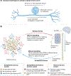
Fig. 1 |
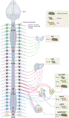
Fig. 1 |. Overview of adipose tissue innervation by sensory and sympathetic nerves and associated release of nerve products (neuropeptides and neurotransmitters).
a, Cartoon of a typical pseudo-unipolar sensory neuron as it innervates adipose tissues. b, Sensory–sympathetic loops between the adipose tissues and the CNS. Sensory nerves (red arrows) stereotypically partake in afferent signalling (as electrical action potentials) from adipose tissue to the CNS via the DRG, and simultaneously release sensory neuropeptides in adipose tissues. Sympathetic nerves (blue arrows) partake in efferent signalling from the CNS to adipose tissues via the sympathetic chain ganglia and release products such as norepinephrine (NE).
Fig. 2 |
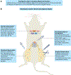
Fig. 2 |. Anatomical distribution of sensory afferent cell bodies across levels of the DRG in rodents (Siberian hamster, mouse and rat) and corresponding peripheral tissue and organ sensory innervation projections.
The DRG houses cell bodies of peripheral sensory nerves that innervate organs and tissues throughout the body (peri-gonadal white adipose tissue (pgWAT) or epididymal WAT (eWAT), peri-renal white adipose tissue (prWAT), BAT and ing-scWAT). For simplicity, the schematic shows only unilateral and not bilateral innervation of organs by sensory afferents. Direction of the arrows shows the direction of sensory neuropeptide release into peripheral organs. DRG levels: C, cervical; T, thoracic; L, lumbar; S, sacral.
Fig. 3 |
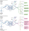
Fig. 3 |. Commonly adopted methods to study adipose innervation in rodents, including adipose denervation techniques.
a, Techniques used to visualize tissue innervation include light-sheet microscopy, whole-mouse or tissue clearing, whole-mount immunofluorescence labelling, intravital imaging and two-photon imaging. b, Methods of achieving sensory denervation of adipose tissues in rodents include chemical denervation, surgical denervation, viral denervation and genetic denervation. 6-OHDA, 6-hydroxydopamine; IB4-Saporin, isolectin B4-Saporin; RTX, resiniferatoxin.
Fig. 4 |
Fig. 4 |. Evidence for positive or negative feedback loops between sensory and sympathetic nerves in adipose tissue.
a, The concept of a positive feedback loop between sensory and sympathetic nerves in adipose tissues is supported by pharmacological agonism or ablation (using high-dose capsaicin) of sensory nerves in ing-scWAT, which measured increases in serum norepinephrine, mean arterial pressure (MAP), norepinephrine turnover (NETO) at room temperature (RT) in BAT, electrophysiological activation of sympathetic nerves in WAT and renal sympathetic nerve activity in the kidneys. b, The concept of a negative feedback loop between sensory and sympathetic nerves in adipose tissue is supported by genetic and viral denervation approaches and is evidenced by the following observations: vasoconstriction with reduced tail temperature at room temperature after systemic genetic ablation of CGRP (Advillin-Cre crossed with Calca-lox-GFP-lox-DTR), increased BAT and core body temperature, and enhanced lipid utilization in BAT after sensory ablation by high-dose capsaicin or systemic genetic CGRP ablation, reduced tyrosine hydroxylase (TH) levels in BAT after orexin receptor agonism in BAT by [Ala11, d-Leu15] OxB, and increased lipid utilization in ing-scWAT after unilateral or bilateral viral sensory denervation with mCherry-flex-DTA injected into T13/L1 DRG with ROOT-Cre injected into ing-scWAT.

References

    1. Bartness TJ, Vaughan CH & Song CK Sympathetic and sensory innervation of brown adipose tissue. Int. J. Obes. 34, S36–S42 (2010). - PMC - PubMed
    1. Bartness TJ, Shrestha YB, Vaughan CH, Schwartz GJ & Song CK Sensory and sympathetic nervous system control of white adipose tissue lipolysis. Mol. Cell. Endocrinol. 318, 34–43 (2010). - PMC - PubMed
    1. Wang Y et al. The role of somatosensory innervation of adipose tissues. Nature 609, 569–574 (2022). - PMC - PubMed
    1. Bartness T & Kay Song C Innervation of brown adipose tissue and its role in thermogenesis. Can. J. Diabetes 29, 420–428 (2005).
    1. Youngstrom TG & Bartness TJ White adipose tissue sympathetic nervous system denervation increases fat pad mass and fat cell number. Am. J. Physiol. 275, R1488–R1493 (1998). - PubMed

Publication types

LinkOut - more resources