

Short-term-outcomes of idiopathic epiretinal membranes treated with pars-plana-vitrectomy - examination of visual function and OCT-morphology
- PMID: 37710332
- PMCID: PMC10500920
- DOI: 10.1186/s40942-023-00496-3
Short-term-outcomes of idiopathic epiretinal membranes treated with pars-plana-vitrectomy - examination of visual function and OCT-morphology
Abstract
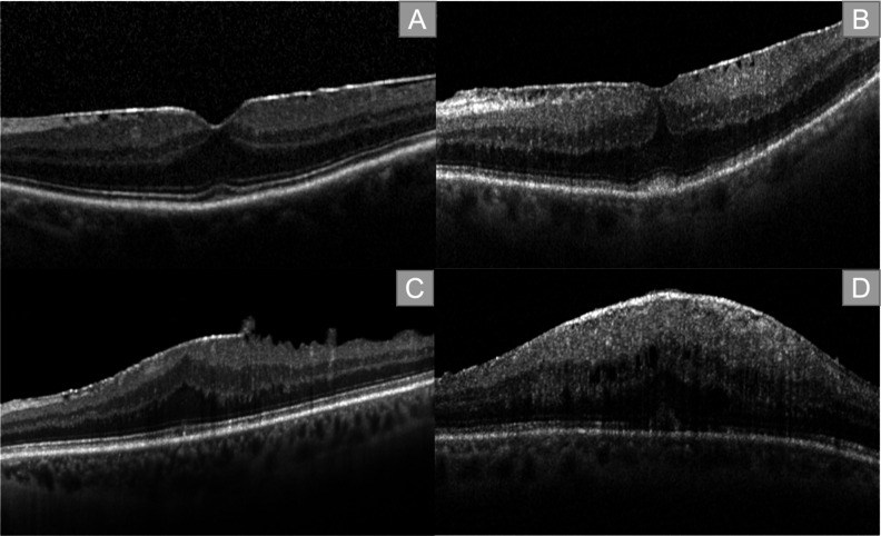
Background: Epiretinal membranes (ERM) represent one of the most common findings in retinal examination. Structural changes of the retinal layers in patients with ERM can be visualized and classified using OCT. The purpose of this study is to evaluate structural and functional changes related to surgical treatment of ERM.
Methods: Monocentric retrospective analysis of 92 patients who underwent 23-gauge-pars plana vitrectomy (ppV) combined with cataract surgery for idiopathic ERM from 2015 to 2020. Visual acuity was determined directly preoperatively, at four weeks and three months postoperatively. Disease stage and tomographic biomarkers related to ERM were assessed in OCT imaging.
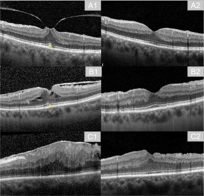
Results: 92 eyes of 92 patients were included. At the time of surgery, the mean patient age was 71 years. Visual acuity improved significantly by 2 lines postoperatively, on average from LogMar 0.4 to 0.2 (p < 0.001). Disease stage regressed from stage 3 to stage 2 postoperatively (p < 0.001). No patient had stage 4 postoperatively (n = 0). In the presence of preoperative intraretinal fluid, mean retinal thickness was 488 μm and decreased to 392 μm postoperatively (n = 32; p < 0.001). Preoperative presence of a Cotton Ball Sign (n = 30) was associated with better visual acuity (p = 0.009). This was also visible in patients with preoperative vitreomacular traction syndrome (p < 0.001). The presence of preoperative intraretinal fluid showed a tendency towards better disease staging after surgery (p = 0.080).
Conclusion: Surgery was able to achieve visual improvement and morphological regression of the preoperative OCT findings related to ERM. ppV led to a reduction in retinal thickness and disease stage. The presence of the Cotton Ball Sign and vitreomacular traction was associated with better visual acuity in the follow-up period. In our cohort the preoperative presence of intraretinal fluid showed a tendency for better postoperative disease staging.
Keywords: Biomarkers; Classification; Gliosis; Oct; Optical coherence tomography; Retina; Staging.
© 2023. Brazilian Retina and Vitreous Society.
Conflict of interest statement
No competing interests can be declared.
Figures
Similar articles
-
Pars Plana Vitrectomy for Idiopathic Epiretinal Membrane: OCT Biomarkers of Visual Outcomes in 322 Eyes.Ophthalmol Retina. 2022 Apr;6(4):308-317. doi: 10.1016/j.oret.2021.10.008. Epub 2021 Oct 27. Ophthalmol Retina. 2022. PMID: 34718218
-
Associations between preoperative OCT parameters and visual outcome 3 months postoperatively in patients undergoing vitrectomy for idiopathic epiretinal membrane.Graefes Arch Clin Exp Ophthalmol. 2016 Oct;254(10):1909-1917. doi: 10.1007/s00417-016-3326-x. Epub 2016 Mar 30. Graefes Arch Clin Exp Ophthalmol. 2016. PMID: 27025926
-
Swept-Source Optical Coherence Tomography Correlations Between Retina and Choroid Before and After Vitrectomy for Epiretinal Membranes.Am J Ophthalmol. 2016 May;165:100-7. doi: 10.1016/j.ajo.2016.02.003. Epub 2016 Mar 10. Am J Ophthalmol. 2016. PMID: 26970574
-
THE EFFECT OF INTERNAL LIMITING MEMBRANE PEELING ON IDIOPATHIC EPIRETINAL MEMBRANE SURGERY, WITH A REVIEW OF THE LITERATURE.Retina. 2017 May;37(5):873-880. doi: 10.1097/IAE.0000000000001263. Retina. 2017. PMID: 27617536 Review.
-
Preoperative ocular coherence tomographic prognosticators of visual acuity after idiopathic epiretinal membrane surgery.Int Ophthalmol. 2022 Oct;42(10):3243-3252. doi: 10.1007/s10792-022-02317-2. Epub 2022 May 18. Int Ophthalmol. 2022. PMID: 35583682 Review.
References
LinkOut - more resources
Full Text Sources
Miscellaneous
