. 2023 Sep 14;13(1):172.
doi: 10.1186/s13578-023-01105-4.

Sex-dependent neuronal effects of α-synuclein reveal that GABAergic transmission is neuroprotective of sleep-controlling neurons

Affiliations

Sex-dependent neuronal effects of α-synuclein reveal that GABAergic transmission is neuroprotective of sleep-controlling neurons

Altair Brito Dos Santos et al. Cell Biosci. .

Abstract

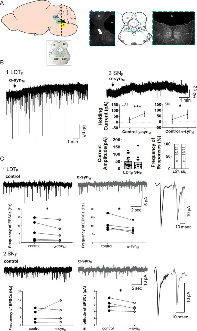
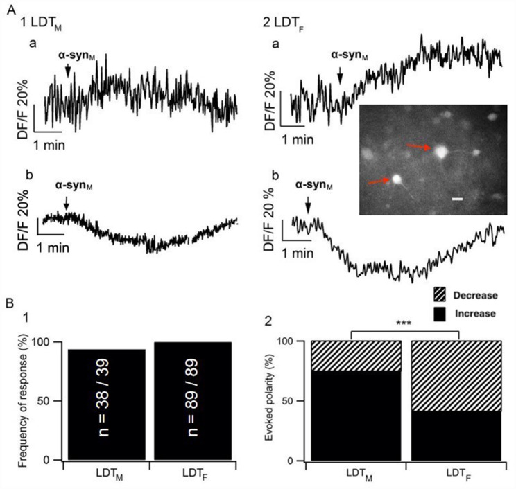
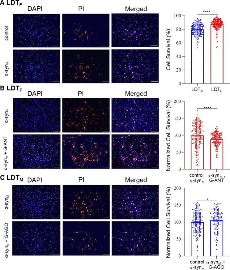
Background: Sleep disorders (SDs) are a symptom of the prodromal phase of neurodegenerative disorders that are mechanistically linked to the protein α-synuclein (α-syn) including Parkinson's disease (PD). SDs during the prodromal phase could result from neurodegeneration induced in state-controlling neurons by accumulation of α-syn predominant early in the disease, and consistent with this, we reported the monomeric form of α-syn (monomeric α-syn; α-synM) caused cell death in the laterodorsal tegmental nucleus (LDT), which controls arousal as well as the sleep and wakefulness state. However, we only examined the male LDT, and since sex is considered a risk factor for the development of α-syn-related diseases including prodromal SDs, the possibility exists of sex-based differences in α-synM effects. Accordingly, we examined the hypothesis that α-synM exerts differential effects on membrane excitability, intracellular calcium, and cell viability in the LDT of females compared to males.

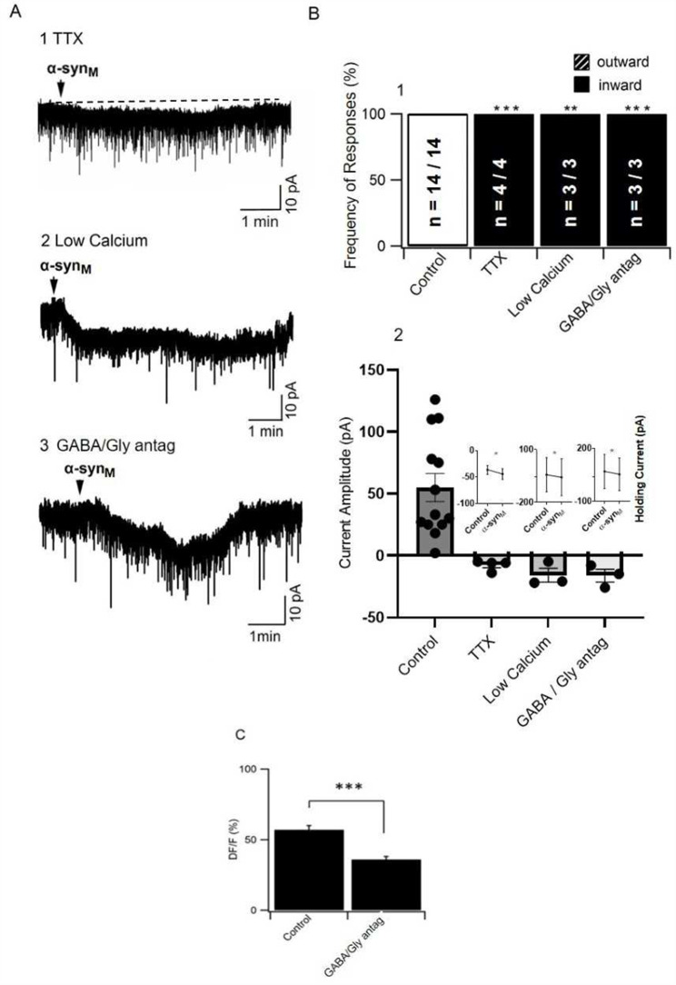
Methods: Patch clamp electrophysiology, bulk load calcium imaging, and cell death histochemistry were used in LDT brain slices to monitor responses to α-synM and effects of GABA receptor acting agents.

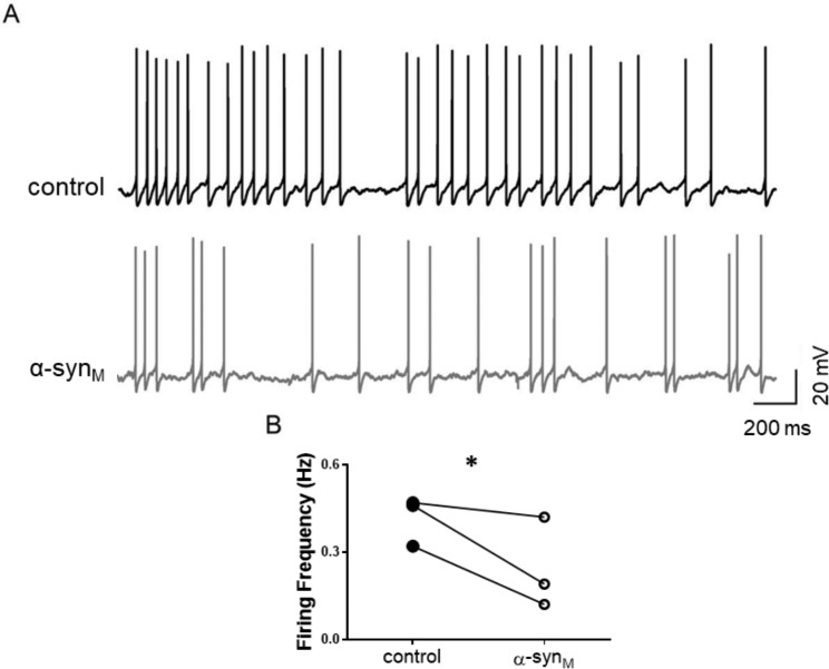
Results: Consistent with our hypothesis, we found differing effects of α-synM on female LDT neurons when compared to male. In females, α-synM induced a decrease in membrane excitability and heightened reductions in intracellular calcium, which were reliant on functional inhibitory acid transmission, as well as decreased the amplitude and frequency of spontaneous excitatory postsynaptic currents (sEPSCs) with a concurrent reduction in action potential firing rate. Cell viability studies showed higher α-synM-mediated neurodegeneration in males compared to females that depended on inhibitory amino acid transmission. Further, presence of GABA receptor agonists was associated with reduced cell death in males.

Conclusions: When taken together, we conclude that α-synM induces a sex-dependent effect on LDT neurons involving a GABA receptor-mediated mechanism that is neuroprotective. Understanding the potential sex differences in neurodegenerative processes, especially those occurring early in the disease, could enable implementation of sex-based strategies to identify prodromal PD cases, and promote efforts to illuminate new directions for tailored treatment and management of PD.

Keywords: Cholinergic; Laterodorsal Tegmentum; Sleep disorders; α -synucleinopathies, neurodegenerative disease.


Conflict of interest statement

All authors disclose that they have no relevant financial or non-financial interests to disclose.

Figures

Fig. 1
Fig. 1
α-synM induced an inhibitory outward current and modulated synaptic transmission in LDT neurons in the female. A) (Left) Cartoon schematic of the sagittal mouse brain to show the block of the brain containing the LDT. A LDT coronal brain slice taken from this block is shown below in inset. (A) (Right) Coronal brain slice cartoon modified from [72] to show in greater detail in insets to the sides the location of the LDT (indicated by white arrow in panel to the left). (B) Sample of membrane response to α-synM, which induced inhibitory, outward currents in the female in the LDT (B1). An outward inhibitory current was also elicited in SN neurons (B2). (B) Graphs of holding currents before and after application of α-synM to LDTF and SNF neurons showed a significant increase in positive holding current indicating that α-synM induced outward currents. The amplitude of the outward current evoked by α-synM in LDTF and SNF neurons was not different (LDTF: n = 14, SNF: n = 11; p = 0.3847; Unpaired Student’s T-test) as shown by the plots of the individual amplitude of current induced in both nuclei. Bar chart showing that the proportion of recorded cells responding to α-synM with induction of outward current did not differ significantly between the LDTF and SNF (LDTF: n = 14 sampled/14 responded, SNF: n = 11 sampled/11 responded; p = 1.000; Fisher’s Exact Test). (C) α-synM modulated synaptic events in neurons recorded within LDTF and SNF. (C1- C2) samples of recordings showing frequency of synaptic events in control and in presence of α-synM in both LDTF and SNF. (Rightmost panels) Single sEPSCs (spontaneous excitatory postsynaptic currents) in a LDTF and in a SNF neuron are shown with a high-gain time and amplitude scale under control conditions and in presence of α-synM illustrating the reduction in amplitude in both nuclei when α-synM was present. Data presented in paired plots summarize findings from the population of recorded cells, which revealed that α-synM induced a significant decrease in amplitude of EPSCs in LDTF (n = 5; p = 0.0253; Paired T-test) and SNF neurons (n = 4; p = 0.0483; Paired T-test) and elicited a significant decrease in the frequency of sEPSCs in LDTF neurons (n = 5; p = 0.0475; Paired T-test), which was a change not seen in sEPSCs in the SN (n = 4; p = 0.4735; Paired T-test). LDT: Laterodorsal tegmental nucleus; 4 V: 4th ventricle; IC: Inferior colliculus; DTgP: Dorsal tegmental nucleus, pericentral: DRN: dorsal raphe nucleus; LC: Locus coeruleus; LDTF: Laterodorsal tegmental nucleus of female; SNF: Substantia nigra of female. * Indicates p < 0.05, *** Indicates p < 0.001
Fig. 2
Fig. 2
Sample of changes in fluorescence (DF/F%) induced by α-synM, which are indicative of alterations in intracellular calcium levels in LDTM and LDTF. (A) In both sexes, changes in response of the fluorescence to α-synM exhibited two different polarities, which suggested increases (A1a, A2a) or decreases (A1b, A2b) in intracellular calcium levels, respectively. Inset in A2 is a fluorescent image under 380 nm wavelength light of one of the LDTF brain slices used in this study in which two Fura 2-AM filled cells indicated with red arrows can be seen. Regions of interest were drawn around each cell and average fluorescent intensity (F) within each region of interest was plotted against time. White scale bar indicates 20 μm. (B) Histograms summarizing the data from the population of recorded cells indicating that (B1) the frequency of responses to α-synM did not significantly differ between the two sexes (LDTM: n = 38/39, LDTF: n = 89/89; p = 0.3047; Fischer’s Exact test), (B2) whereas the distribution of response polarity differed significantly between the sexes with a greater proportion of responses suggesting decreases in calcium being elicited in females than males (LDTM: n of decreases = 9/38, LDTF: n of decreases = 52/89; p = 0.0004; Fisher’s Exact test). LDTM: Laterodorsal tegmental nucleus of male; LDTF: Laterodorsal tegmental nucleus of female. *** Indicates p < 0.001
Fig. 3
Fig. 3
An excitatory inward current was revealed when α-synM was applied during blockade of presynaptic transmission, or antagonism of GABAA, GABAB and glycine receptors, suggesting that α-synM induces an outward membrane current in LDTF neurons due to actions at presynaptic inhibitory neurons. A) Sample of membrane responses to α-synM in which an inward current is revealed when α-synM is applied in presence of (A1) TTX, (A2) low calcium solution, or (A3) a cocktail of SR-95,531 + CGP-55,845 + strychnine, which block GABAA, GABAB and glycine receptors, respectively). (B) Histograms from a population of cells recorded in which there were significant changes in the (B1) polarity of the evoked membrane current responses to α-synM in presence of TTX, low calcium solution or GABA and glycine receptor antagonists when compared to control responses (Fisher’s Exact test). (B2) The amplitude of the current evoked by α-synM in control conditions and under conditions of synaptic blockade and inhibitory receptor antagonists is shown revealing the change in polarity of the α-synM induced current. (B2, Inset) Graphs of holding currents before and after application of α-synM to LDTF showed that in presence of TTX, low calcium solution, and GABAA, GABAB, and glycine receptor antagonists, holding currents became significantly more negative after application of α-synM, which reflected the α-synM-mediated induction of inward currents. (C) The decrease in intracellular calcium in LDTF is mediated, at least in part, by inhibitory receptors as illustrated in this histogram showing data from the population of LDTF cells recorded that showed a significantly smaller decrease in the amplitude of intracellular calcium induced by α-synM in presence of SR-95,531, CGP-55,845 and strychnine when compared to control conditions (Control: n = 52, GABA/Gly antagonists: n = 49; p = 0.0001; Paired T-test). LDTF: Laterodorsal tegmental nucleus of female. * Indicates p < 0.05, ** Indicates p < 0.01, *** Indicates p < 0.001
Fig. 4
Fig. 4
α-synM induces significant changes in the firing frequency in neurons recorded within LDTF. (A) Representative examples of current-clamp recordings of LDT neurons in which action potentials were induced by holding the cell at -45 mV under control conditions (top) and in presence of α-synM. (bottom). (B) The reduction in firing frequency induced by α-synM was significant as shown in the bar graphs depicting the average firing rate from the population of recorded LDTF neurons (n = 3; p = 0.0498; Paired T-test). LDTF: Laterodorsal tegmental nucleus of female * Indicates p < 0.05
Fig. 5
Fig. 5
(A-C, left panels) DAPI and PI immunohistochemistry conducted in LDTF and LDTM from brain slices exposed to α-synM under 3 different treatment protocols is shown in representative fluorescent images. The first column represents living cells visualized by DAPI (blue). The second column represents dead cells visualized by PI (red), and the last column is a merged image of DAPI and PI labeled cells. (A) The presence of DAPI and PI in the LDTF following incubation of one half of a slice in control solution (ACSF) and the other half in ACSF containing α-synM for 7 h is shown and indicates relatively lower cell survival in the half of the slice exposed to α-synM, which was reflected in the population data. The bar graph to the right shows that following α-synM exposure, cell survival in LDTF was significantly greater than that seen in LDTM (Cell Survival Female: na = 183/n40 = 48, Cell Survival Male: na=168/n40 = 66; p < 0.0001; Unpaired Student’s T-test). In this and subsequent panels, red points represent observations from LDTF, blue represent data from LDTM, and cell counts within each area (na) represent one data point or observation in the bar chart columns. (B) Fluorescent images showing DAPI and PI presence in LDTF cells treated for 7 h with α-synM or with α-synM in presence of GABAA, GABAB and glycine receptors antagonists (G-ANT). As shown in the bar graph to the right, a reduced cell survival indicative of greater cell mortality was observed in the population of LDTF slices exposed to α-synM when GABA and glycine receptor antagonists were present (Cell survival: α-synM: na = 199/n40 = 44, Cell survival α-synM + G-ANT: na = 169/n40 = 35; p = 0.0001; Mann-Whitney Test). To compare the population data, bisected slices were used, and the proportion of surviving cells observed in the half of the bisected slice exposed to α-synM was considered the baseline, and the number of surviving cells in the other half of the bisected slice exposed to α-synM + G-ANT was normalized to this baseline. (C) Fluorescent images of LDTM slices exposed to ⍺-synM or to α-synM in presence of 7 h of GABAA and GABAB receptor agonists (G-AGO). As can be seen from the population data shown in bar graphs to the right, the presence of the GABA and glycine receptor agonists in the LDTM was associated with significantly greater cell survival following exposure to α-synM (Cell Survival α-synM: na = 168/n40 = 66, Cell Survival α-synM + G-AGO: na = 127 /n40 = 48; p = 0.0334; Mann-Whitney Test). In this protocol, the proportion of surviving cells observed in the half of the bisected slice exposed to α-synM was considered the baseline, and the number of surviving cells in the other half of the bisected slice exposed to α-synM + G-AGO was normalized to this baseline. LDTF: the laterodorsal tegmental nucleus of female; LDTM: the laterodorsal tegmental nucleus of male. G-ANT: contains SR-95,531 (gabazine, 10 µM), CGP 55,845 (10 µM) and strychnine (2.5 µM) to block GABAA, GABAB, and glycine receptor-mediated responses, respectively. G-AGO: contains muscimol (30 µM) and baclofen (10 µM), which are agonists of GABAA and GABAB receptors, respectively. The scale bar in all images corresponds to 50 μm under 40x magnification. Contrast has been added equally across all the images. *p < 0.05, **** p < 0.0001

Similar articles

Cited by

References

    1. Goetz CG. The history of Parkinson’s Disease: early clinical descriptions and neurological therapies, Cold Spring Harb. Perspect Med. 2011;1:a008862–2. doi: 10.1101/cshperspect.a008862. - DOI - PMC - PubMed
    1. Fahn S. Description of Parkinson’s Disease as a clinical syndrome. Ann N Y Acad Sci. 2006;991:1–14. doi: 10.1111/j.1749-6632.2003.tb07458.x. - DOI - PubMed
    1. Poewe W, Seppi K, Tanner CM, Halliday GM, Brundin P, Volkmann J, Schrag A-E, Lang AE. Parkinson disease. Nat Rev Dis Prim. 2017;3:17013. doi: 10.1038/nrdp.2017.13. - DOI - PubMed
    1. Elbaz A, Bower JH, Maraganore DM, McDonnell SK, Peterson BJ, Ahlskog JE, Schaid DJ, Rocca WA. Risk tables for parkinsonism and Parkinson’s disease. J Clin Epidemiol. 2002;55:25–31. doi: 10.1016/S0895-4356(01)00425-5. - DOI - PubMed
    1. de Lau LML, Giesbergen PCLM, de Rijk MC, Hofman A, Koudstaal PJ, Breteler MMB. Incidence of parkinsonism and Parkinson disease in a general population: the Rotterdam Study, Neurology. 63 (2004) 1240–4. 10.1212/01.WNL.0000140706.52798.BE. - PubMed

LinkOut - more resources