

Pharmacological elevation of sphingosine-1-phosphate by S1P lyase inhibition accelerates bone regeneration after post-traumatic osteomyelitis
- PMID: 37710406
- PMCID: PMC10718149
- DOI: 10.1111/jcmm.17952
Pharmacological elevation of sphingosine-1-phosphate by S1P lyase inhibition accelerates bone regeneration after post-traumatic osteomyelitis
Abstract
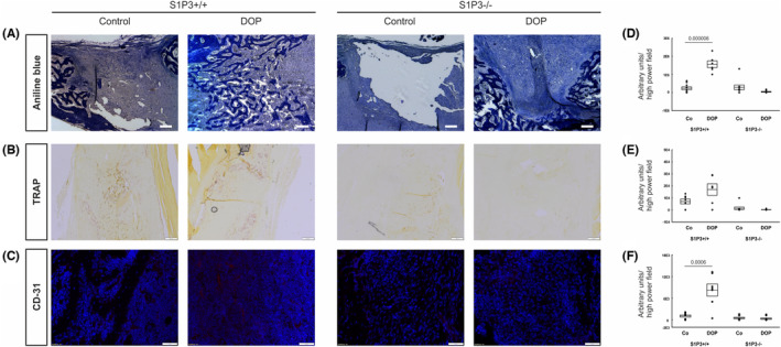
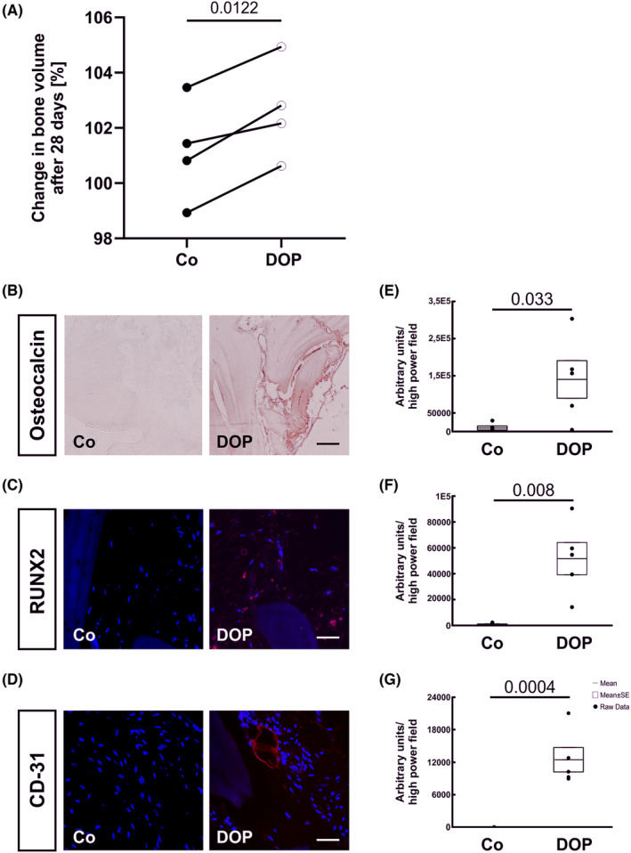
Posttraumatic osteomyelitis and the ensuing bone defects are a debilitating complication after open fractures with little therapeutic options. We have recently identified potent osteoanabolic effects of sphingosine-1-phosphate (S1P) signalling and have now tested whether it may beneficially affect bone regeneration after infection. We employed pharmacological S1P lyase inhibition by 4-deoxypyrodoxin (DOP) to raise S1P levels in vivo in an unicortical long bone defect model of posttraumatic osteomyelitis in mice. In a translational approach, human bone specimens of clinical osteomyelitis patients were treated in organ culture in vitro with DOP. Bone regeneration was assessed by μCT, histomorphometry, immunohistology and gene expression analysis. The role of S1P receptors was addressed using S1PR3 deficient mice. Here, we present data that DOP treatment markedly enhanced osteogenesis in posttraumatic osteomyelitis. This was accompanied by greatly improved osteoblastogenesis and enhanced angiogenesis in the callus accompanied by osteoclast-mediated bone remodelling. We also identified the target of increased S1P to be the S1PR3 as S1PR3-/- mice showed no improvement of bone regeneration by DOP. In the human bone explants, bone mass significantly increased along with enhanced osteoblastogenesis and angiogenesis. Our data suggest that enhancement of S1P/S1PR3 signalling may be a promising therapeutic target for bone regeneration in posttraumatic osteomyelitis.
Keywords: bone regeneration; osteomyelitis; sphingosin-1-phosphate.
© 2023 The Authors. Journal of Cellular and Molecular Medicine published by Foundation for Cellular and Molecular Medicine and John Wiley & Sons Ltd.
Conflict of interest statement
The authors declare they have no conflict of interest.
Figures


Similar articles
-
Sphingosine-1-phosphate promotes osteogenesis by stimulating osteoblast growth and neovascularization in a vascular endothelial growth factor-dependent manner.J Bone Miner Res. 2024 Apr 19;39(3):357-372. doi: 10.1093/jbmr/zjae006. J Bone Miner Res. 2024. PMID: 38477738 Free PMC article.
-
HuR mediates motility of human bone marrow-derived mesenchymal stem cells triggered by sphingosine 1-phosphate in liver fibrosis.J Mol Med (Berl). 2017 Jan;95(1):69-82. doi: 10.1007/s00109-016-1460-x. Epub 2016 Aug 20. J Mol Med (Berl). 2017. PMID: 27543493
-
Increased S1P expression in osteoclasts enhances bone formation in an animal model of Paget's disease.J Cell Biochem. 2021 Apr;122(3-4):335-348. doi: 10.1002/jcb.29861. Epub 2020 Oct 27. J Cell Biochem. 2021. PMID: 33107091 Free PMC article.
-
Sphingosine 1-phosphate (S1P) signalling: Role in bone biology and potential therapeutic target for bone repair.Pharmacol Res. 2017 Nov;125(Pt B):232-245. doi: 10.1016/j.phrs.2017.08.013. Epub 2017 Sep 22. Pharmacol Res. 2017. PMID: 28855094 Free PMC article. Review.
-
Sphingosine 1-phosphate signaling in bone remodeling: multifaceted roles and therapeutic potential.Expert Opin Ther Targets. 2017 Jul;21(7):725-737. doi: 10.1080/14728222.2017.1332180. Epub 2017 Jun 7. Expert Opin Ther Targets. 2017. PMID: 28524744 Free PMC article. Review.
References
Publication types
MeSH terms
Substances
Grants and funding
LinkOut - more resources
Full Text Sources
Other Literature Sources
