. 2023 Nov;46(6):1195-1205.
doi: 10.1002/jimd.12679. Epub 2023 Oct 5.

Biallelic missense variants in COG3 cause a congenital disorder of glycosylation with impairment of retrograde vesicular trafficking

Affiliations

Biallelic missense variants in COG3 cause a congenital disorder of glycosylation with impairment of retrograde vesicular trafficking

Ruizhi Duan et al. J Inherit Metab Dis. 2023 Nov.

Abstract

Biallelic variants in genes for seven out of eight subunits of the conserved oligomeric Golgi complex (COG) are known to cause recessive congenital disorders of glycosylation (CDG) with variable clinical manifestations. COG3 encodes a constituent subunit of the COG complex that has not been associated with disease traits in humans. Herein, we report two COG3 homozygous missense variants in four individuals from two unrelated consanguineous families that co-segregated with COG3-CDG presentations. Clinical phenotypes of affected individuals include global developmental delay, severe intellectual disability, microcephaly, epilepsy, facial dysmorphism, and variable neurological findings. Biochemical analysis of serum transferrin from one family showed the loss of a single sialic acid. Western blotting on patient-derived fibroblasts revealed reduced COG3 and COG4. Further experiments showed delayed retrograde vesicular recycling in patient cells. This report adds to the knowledge of the COG-CDG network by providing collective evidence for a COG3-CDG rare disease trait and implicating a likely pathology of the disorder as the perturbation of Golgi trafficking.

Keywords: AOH/ROH analysis; congenital disorders of glycosylation; conserved oligomeric Golgi complex; family-based genomic analysis; retrograde vesicular transport.


Conflict of interest statement

Conflict of Interest

James R. Lupski has stock ownership in 23andMe, is a paid consultant for Genomics International, and is a co-inventor on multiple United States and European patents related to molecular diagnostics for inherited neuropathies, eye diseases, genomic disorders, and bacterial genomic fingerprinting. The Department of Molecular and Human Genetics at Baylor College of Medicine receives revenue from clinical genetic testing conducted at Baylor Genetics (BG); James R. Lupski serves on the Scientific Advisory Board (SAB) of BG. Ruizhi Duan, Dana Marafi1, Zhi-Jie Xia, Bobby G. Ng, Reza Maroofian, Farhana Taher Sumya, Ahmed K. Saad, Haowei Du, Jawid M. Fatih, Jill V. Hunter, Hasnaa M. Elbendary, Shahid M. Baig, Uzma Abdullah, Zafar Ali, Stephanie Efthymiou, David Murphy, Tadahiro Mitani, Marjorie A. Withers, Shalini N. Jhangiani, Zeynep Coban-Akdemir, Daniel G. Calame, Davut Pehlivan, Richard A. Gibbs, Jennifer E. Posey, Henry Houlden, Vladimir V. Lupashin, Maha S. Zaki and Hudson H. Freeze declare that they have no conflict of interest.

Figures
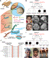
Figure 1.
Figure 1.
Comprehensive illustration, familial pedigree, clinical photographs and genetic findings of the identified homozygous variant in COG3 (A) An illustrative graph briefly describes the of COG-mediated vesicular tethering. During the early phase of membrane fusion, partially coated vesicle can be captured by the of multisubunit tethering machinery including the COG complex (as the recognitional bridge), soluble N-ethylmaleimide-sensitive factor attachment protein receptor (SNARE) and Rab GTPases. Structural diagram of COG Lobe-A was adapted from the publication (Lee et al, Nat Struct Mol Biol, 2010). The reported variant p.(Ser42Pro) resides in the COG3 N-terminus, a long segment of α-helix presumably involved quaternary interactions with the other three subunits (shaded by blue rectangle in the diagram). Location of the variant was highlighted by 3D protein structural modeling of COG3 and COG4 obtained from AlphaFold Protein Structure Database (https://alphafold.ebi.ac.uk/). (B) Pedigree and allelic information with the segregation of the missense variant COG3 [NM_031431.4: c.124T>C, p.(Ser42Pro)] in the family 1 (HOU4978). (C) Clinical photograph of the female affected individual BAB13440 showing mild facial dysmorphism. (D-E) Brain MRI (Axial and midsagittal T2 weighted imaging, respectively) of BAB13340 at 5 years of age showing mild right posterior plagiocephaly, generous CSF spaces, wide interhemispheric fissure, significant cerebral volume loss, bilateral symmetric under-opercularization, delayed myelination for age, thinning of corpus callosum, prominent supratentorial and fourth ventricles, and mild superior vermian hypoplasia. The findings reflect a more severe supratentorial than infratentorial involvement. (F) Clinical photograph of the deceased individual BAB13337 showing mild facial dysmorphism and strabismus (G-H) Brain MRI (Axial and midsagittal T2 weight imaging, respectively) BAB13337 at 4 months of age showing right posterior plagiocephaly, generous CSF spaces, prominent cerebral volume loss, bilateral symmetric under-opercularization, delayed myelination for age, thinning of corpus callosum, normal supratentorial ventricular system with large 4th ventricle, and very mild superior vermian hypoplasia. (I) Pedigree and allelic information with the segregation of the missense variant COG3 [NM_031431.4: c.109G>C, p.(Asp37His)] in family 2 (MR-176). Note: the there are two deceased affected female individuals in this family. The elder one (SYNS-01449) was confirmed harboring the homozygous variant of COG3 with phenotype information reported. While the genotype and phenotype information of the younger deceased individual (female, affected) is not available. (J) The altered amino acids and other adjacent residues are evolutionarily conserved. (K) A clinical photograph of the proband SYNS-01448 shows subtle facial dysmorphism. (L) A photograph of the deceased female affected individual SYNS-01449 shows a similar facial feature. (M) A photograph of the proband SYNS-01448 shows muscle wasting in the legs.
Figure 2.
Figure 2.
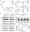
Reduced expression of COG3 and COG4 with delayed retrograde trafficking (A) Biochemical analysis on serum transferrin from two affected individuals in family 1 showed aberrant sialyation with a higher ratio of ‘tri-sialo/di-oligo’ compared with a healthy individual control via MAYO electrospray ionization mass spectrometry. (B-C) Western blot images of COG3, COG1, COG2 and COG4 abundance in unaffected control, BAB13339, and BAB13340 fibroblasts. Relative COG3 level was normalized to β-actin, the internal loading control of Western blotting. The experiment was performed in duplicate with similar results. The reduction of COG3 was seen in BAB13339 by more than 50% compared to unaffected controls. A significantly reduced COG4 was seen in BAB13339 compared to unaffected controls. The experiment was performed in duplicate with similar results. Unpaired two-tailed t-test was used. *, p<0.05; **, p<0.01. (D) BFA-induced retrograde transport of Giantin in unaffected controls, BAB13339, BAB13340, and other COG-CDG individuals’ fibroblasts. The percentage of ER staining pattern of Giantin was calculated at 0, 5, 10, 15, 22.5, and 30 min with BFA treatment in fixed samples. 80–100 cells were counted at each time point. Experiments were done in triplicate. COG4, LoF COG4-CDG individual’s fibroblast. COG8, COG8-CDG individual’s fibroblast.

References

    1. Park S-Y, Yang J-S, Schmider AB, Soberman RJ, Hsu VW. Coordinated regulation of bidirectional COPI transport at the Golgi by CDC42. Nature. 2015;521(7553):529–532. doi:10.1038/nature14457 - DOI - PMC - PubMed
    1. Nakano A Polarized transport in the Golgi apparatus. Nature. 2015;521(7553):427–428. doi:10.1038/nature14521 - DOI - PubMed
    1. Reily C, Stewart TJ, Renfrow MB, Novak J. Glycosylation in health and disease. Nat Rev Nephrol. 2019;15(6):346–366. doi:10.1038/s41581-019-0129-4 - DOI - PMC - PubMed
    1. Freeze HH, Chong JX, Bamshad MJ, Ng BG. Solving glycosylation disorders: Fundamental approaches reveal complicated pathways. Am J Hum Genet. 2014;94(2):161–175. doi:10.1016/j.ajhg.2013.10.024 - DOI - PMC - PubMed
    1. Climer LK, Dobretsov M, Lupashin V. Defects in the COG complex and COG-related trafficking regulators affect neuronal Golgi function. Front Neurosci. 2015;9(OCT):1–9. doi:10.3389/fnins.2015.00405 - DOI - PMC - PubMed

Publication types

MeSH terms

Substances