

The neurophysiological basis of stress and anxiety - comparing neuronal diversity in the bed nucleus of the stria terminalis (BNST) across species
- PMID: 37711509
- PMCID: PMC10499361
- DOI: 10.3389/fncel.2023.1225758
The neurophysiological basis of stress and anxiety - comparing neuronal diversity in the bed nucleus of the stria terminalis (BNST) across species
Abstract
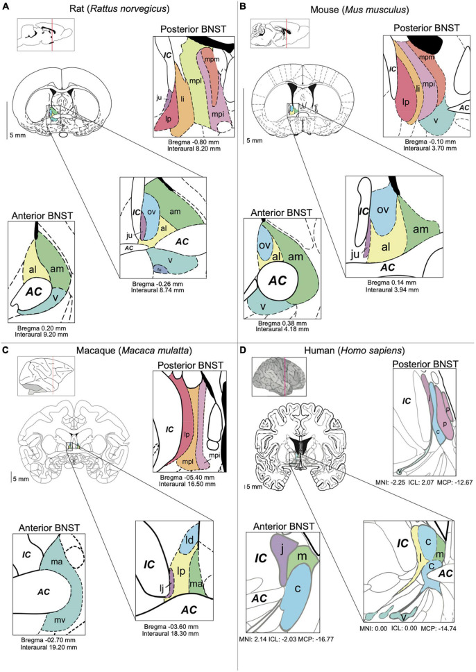
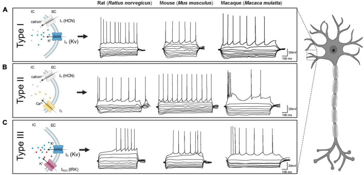
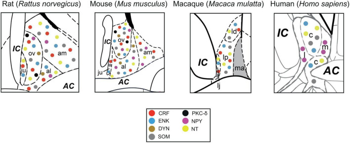
The bed nucleus of the stria terminalis (BNST), as part of the extended amygdala, has become a region of increasing interest regarding its role in numerous human stress-related psychiatric diseases, including post-traumatic stress disorder and generalized anxiety disorder amongst others. The BNST is a sexually dimorphic and highly complex structure as already evident by its anatomy consisting of 11 to 18 distinct sub-nuclei in rodents. Located in the ventral forebrain, the BNST is anatomically and functionally connected to many other limbic structures, including the amygdala, hypothalamic nuclei, basal ganglia, and hippocampus. Given this extensive connectivity, the BNST is thought to play a central and critical role in the integration of information on hedonic-valence, mood, arousal states, processing emotional information, and in general shape motivated and stress/anxiety-related behavior. Regarding its role in regulating stress and anxiety behavior the anterolateral group of the BNST (BNSTALG) has been extensively studied and contains a wide variety of neurons that differ in their electrophysiological properties, morphology, spatial organization, neuropeptidergic content and input and output synaptic organization which shape their activity and function. In addition to this great diversity, further species-specific differences are evident on multiple levels. For example, classic studies performed in adult rat brain identified three distinct neuron types (Type I-III) based on their electrophysiological properties and ion channel expression. Whilst similar neurons have been identified in other animal species, such as mice and non-human primates such as macaques, cross-species comparisons have revealed intriguing differences such as their comparative prevalence in the BNSTALG as well as their electrophysiological and morphological properties, amongst other differences. Given this tremendous complexity on multiple levels, the comprehensive elucidation of the BNSTALG circuitry and its role in regulating stress/anxiety-related behavior is a major challenge. In the present Review we bring together and highlight the key differences in BNSTALG structure, functional connectivity, the electrophysiological and morphological properties, and neuropeptidergic profiles of BNSTALG neurons between species with the aim to facilitate future studies of this important nucleus in relation to human disease.
Keywords: bed nucleus of the stria terminalis (BNST); cross-species; electrophysiology; human; macaque; neurpeptides; rodents.
Copyright © 2023 van de Poll, Cras and Ellender.
Conflict of interest statement
The authors declare that the research was conducted in the absence of any commercial or financial relationships that could be construed as a potential conflict of interest.
Figures
References
-
- Ahearn E. P., Juergens T., Smith T., Krahn D., Kalin N. (2007). Fear, anxiety, and the neuroimaging of PTSD. Psychopharmacol. Bull. 40 88–103. - PubMed
-
- Alexander M. J., Miller M. A., Dorsa D. M., Bullock B. P., Melloni R., Dobner P. R., et al. (1989). Distribution of neurotensin/neuromedin N mRNA in rat forebrain: Unexpected abundance in hippocampus and subiculum. Proc. Natl. Acad. Sci. U. S. A. 86 5202–5206. 10.1073/pnas.86.13.5202 - DOI - PMC - PubMed
Publication types
LinkOut - more resources
Full Text Sources