. 2023 Aug 30:14:1238297.
doi: 10.3389/fimmu.2023.1238297. eCollection 2023.

Murine model identifies tropomyosin as IgE cross-reactive protein between house dust mite and coho salmon that possibly contributes to the development of salmon allergy

Affiliations

Murine model identifies tropomyosin as IgE cross-reactive protein between house dust mite and coho salmon that possibly contributes to the development of salmon allergy

Risa Yamamoto et al. Front Immunol. .

Abstract

Background: Recently, we have developed a method to identify IgE cross-reactive allergens. However, the mechanism by which IgE cross-reactive allergens cause food allergy is not yet fully understood how. In this study, we aimed to understand the underlying pathogenesis by identifying food allergens that cross-react with house dust mite allergens in a murine model.

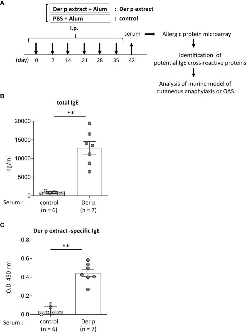
Material and methods: Allergenic protein microarray analysis was conducted using serum from mice intraperitoneally injected with Dermatophagoides pteronyssinus (Der p) extract plus alum or alum alone as controls. Der p, Dermatophagoides farinae (Der f), coho salmon extract-sensitized and control mice were analyzed. Serum levels of IgE against Der p, Der f, coho salmon extract, protein fractions of coho salmon extract separated by ammonium sulfate precipitation and anion exchange chromatography, and recombinant coho salmon tropomyosin or actin were measured by an enzyme-linked immunosorbent assay. A murine model of cutaneous anaphylaxis or oral allergy syndrome (OAS) was established in Der p extract-sensitized mice stimulated with coho salmon extract, tropomyosin, or actin.

Results: Protein microarray analysis showed that coho salmon-derived proteins were highly bound to serum IgE in Der p extract-sensitized mice. Serum IgE from Der p or Der f extract-sensitized mice was bound to coho salmon extract, whereas serum IgE from coho salmon extract-sensitized mice was bound to Der p or Der f extract. Analysis of the murine model showed that cutaneous anaphylaxis and oral allergic reaction were evident in Der p extract-sensitized mice stimulated by coho salmon extract. Serum IgE from Der p or Der f extract-sensitized mice was bound strongly to protein fractions separated by anion exchange chromatography of coho salmon proteins precipitated with 50% ammonium sulfate, which massively contained the approximately 38 kDa protein. We found that serum IgE from Der p extract-sensitized mice was bound to recombinant coho salmon tropomyosin. Der p extract-sensitized mice exhibited cutaneous anaphylaxis in response to coho salmon tropomyosin.

Conclusion: Our results showed IgE cross-reactivity of tropomyosin between Dermatophagoides and coho salmon which illustrates salmon allergy following sensitization with the house dust mite Dermatophagoides. Our method for identifying IgE cross-reactive allergens will help understand the underlying mechanisms of food allergies.

Keywords: IgE cross-reactivity; food allergy; house dust mite; mast cell; murine model; salmon.


Conflict of interest statement

The authors declare that the research was conducted in the absence of any commercial or financial relationships that could be construed as a potential conflict of interest.

Figures

Figure 1
Figure 1
Identification of foods that may cross-react with Der p extract by using a murine model of sensitization with Der p extract and allergenic protein microarray technology.(A) Allergenic protein microarray analysis was performed using serum from BALB/c mice that had been intraperitoneally injected with Der p extract plus alum (Der p extract) or PBS plus alum as a control (control) six times at a 1- week interval, prior to analysis of the murine model of cutaneous anaphylaxis or OAS. (B, C) Serum levels of total IgE (B) and Der p extract-specific IgE (C) from mice on day 42. Data are representative of two independent experiments. Means ± SD have been plotted. **P < 0.01.
Figure 2
Figure 2
Serum IgE from mice sensitized with the Der p extract was significantly bound to the salmon extract. (A) A schematic representation of mouse sensitization with Der p extract. Mice were intraperitoneally injected with Der p extract plus alum or PBS plus alum alone followed by intranasal administration of Der p extract or PBS to generate Der p extract-sensitized mice (Der p extract) or control mice (control), respectively. On day 35, blood samples were obtained. (B, D) Serum levels of specific IgE from Der p extract-sensitized mice or control mice against (B) Der p extract and (D) coho salmon extract. (C, E) Serum levels of specific IgE from Der p extract-sensitized mice or control mice against (D) Der p extract and (F) coho salmon extract after serum preincubation with Der p extract or PBS as a control. (B–E) Data were pooled from three independent experiments. Means ± SD have been plotted. **P < 0.01.
Figure 3
Figure 3
Serum IgE from mice sensitized with coho salmon extract was significantly bound to Der p and Der f extract. (A) A schematic representation of mouse sensitization with Der p or Der f extract. Mice were intraperitoneally injected with Der p or Der f extract plus alum or PBS plus alum alone followed by intranasal administration of Der p or Der f extract or PBS to generate Der p or Der f extract-sensitized mice (Der p extract or Der p extract) or control mice (control), respectively. On day 35, blood samples were obtained. (B) Serum levels of specific IgE from Der p or Der f extract-sensitized mice or control mice against Der p extract (left), Der f extract (middle), or coho salmon extract (right). (C) A schematic representation of mouse sensitization with coho salmon extract. Mice were intraperitoneally injected with coho salmon extract plus alum or PBS plus alum six times at a 1- week interval to generate coho salmon extract-sensitized mice (salmon extract) or control mice (control), respectively. On day 42, blood samples were obtained. (D) Serum levels of specific IgE from salmon extract-sensitized mice or control mice against Der p extract (left), Der f extract (middle), or coho salmon extract (right). (B, D) Data are representative of two independent experiments. Means ± SD have been plotted. *P < 0.05 or **P < 0.01.
Figure 4
Figure 4
Both cutaneous anaphylaxis and oral allergy were evident in Der p extract-sensitized mice after administration of coho salmon extract. (A) A schematic representation of the murine model of cutaneous anaphylaxis or OAS. Mice were intraperitoneally injected with Der p extract plus alum or PBS plus alum followed by intranasal administration of Der p extract or PBS to generate Der p extract-sensitized mice (Der p extract) or control mice (control), respectively. On day 35, mice were challenged with coho salmon extract. (B) The frequency of sneezing during 30 min after nasal administration of Der p extract or PBS on day 32. (C–E) The ear skin was removed from Der p extract-sensitized mice or control mice stimulated by coho salmon extract in murine model of cutaneous anaphylaxis. (C) Quantification of the Evans blue dye. (D) Percentages of degranulated mast cells. (E) Representative images of chloroacetate esterase-stained mast cells in tissue sections. The arrowhead indicates the degranulated mast cell. (F) Quantification of the Evans blue dye that extravasated into the neck skin from Der p extract-sensitized mice or control mice stimulated by coho salmon extract in the murine model of OAS. (B–F) Data are representative of two independent experiments. Means ± SD have been plotted. *P < 0.05 or **P < 0.01.
Figure 5
Figure 5
The molecular weight of the approximately 38-kDa protein included in coho salmon extract was possibly recognized by serum IgE from Der p extract-sensitized mice. (A) Serum levels of specific IgE from Der p or Der f extract-sensitized mice (Der p extract or Der f extract), control mice (control), or coho salmon extract-sensitized mice (salmon extract) against coho salmon proteins precipitated with 30%, 40%, 50%, 60%, or 70% ammonium sulfate (30% ASP, 40% ASP, 50% ASP, 60% ASP, 70% ASP) or soluble in 70% ammonium sulfate (70% ASP supernatant). (B) Coho salmon proteins precipitated with 50% ammonium sulfate were subjected to anion exchange chromatography. Spectra of anion exchange chromatography fractions are shown. The blue line indicates a UV 280 nm signal. The green line indicates NaCl concentration in the buffer. (C) Equivalent amounts of total protein in each fraction of A2, A3, and C7 to C14 were subjected to SDS-PAGE before CBB staining. (D) Serum levels of specific IgE from Der p extract-sensitized mice or control mice against proteins of each fraction (A2, A3, and C7 to C14), salmon extracts, or vehicle (PBS). (A–D) Data are representative of two independent experiments. (A) Means ± SD have been plotted. *P < 0.05 or **P < 0.01. ns, not significant.
Figure 6
Figure 6
Tropomyosin was identified as IgE cross-reactive protein between Der p and coho salmon extract. (A) Serum levels of specific IgE from Der p extract-sensitized mice (Der p extract) or control mice (control) against recombinant coho salmon tropomyosin or actin, coho salmon extracts, or vehicle (PBS). (B) Serum levels of specific IgE from Der p extract-sensitized mice after serum preincubation with coho salmon extract, recombinant coho salmon tropomyosin, or PBS. (C) Percentages of amino acid sequence identity of Der p 10/tropomyosin from American house dust mite with Der f 10/tropomyosin from European house dust mite, tropomyosin 1 from Homarus americanus (American lobster), tropomyosin 1 from Oncorhynchus kisutch (coho salmon), tropomyosin α-1 from Mus musculus (mouse), or tropomyosin α-1 from Homo sapiens (human). The accession number (UniProt) is shown. (D) Quantification of the Evans blue dye that extravasated into the ear skin from Der p extract-sensitized mice or control mice stimulated by coho salmon proteins precipitated with 50% ammonium sulfate (left panel) or recombinant coho salmon tropomyosin or actin (right panel) in murine model of cutaneous anaphylaxis. (A, B, D) Data are representative of two independent experiments. Means ± SD have been plotted. *P < 0.05 or **P < 0.01.

References

    1. Valenta R, Hochwallner H, Linhart B, Pahr S. Food allergies: the basics. Gastroenterology (2015) 148(6):1120–31.e4. doi: 10.1053/j.gastro.2015.02.006 - DOI - PMC - PubMed
    1. Nakano N, Kitaura J. Mucosal mast cells as key effector cells in food allergies. Cells (2022) 11(3):329. doi: 10.3390/cells11030329 - DOI - PMC - PubMed
    1. Kitaura J, Murakami M. Positive and negative roles of lipids in mast cells and allergic responses. Curr Opin Immunol (2021) 72:186–95. doi: 10.1016/j.coi.2021.06.001 - DOI - PubMed
    1. Oyoshi MK, Oettgen HC, Chatila TA, Geha RS, Bryce PJ. Food allergy: Insights into etiology, prevention, and treatment provided by murine models. J Allergy Clin Immunol (2014) 133(2):309–17. doi: 10.1016/j.jaci.2013.12.1045 - DOI - PMC - PubMed
    1. Tordesillas L, Berin MC, Sampson HA. Immunology of food allergy. Immunity (2017) 47(1):32–50. doi: 10.1016/j.immuni.2017.07.004 - DOI - PubMed

Publication types