. 2023 Aug 30:14:1253568.
doi: 10.3389/fimmu.2023.1253568. eCollection 2023.

LAMP1 targeting of the large T antigen of Merkel cell polyomavirus results in potent CD4 T cell responses and tumor inhibition

Affiliations

LAMP1 targeting of the large T antigen of Merkel cell polyomavirus results in potent CD4 T cell responses and tumor inhibition

Claire Buchta Rosean et al. Front Immunol. .

Abstract

Introduction: Most cases of Merkel cell carcinoma (MCC), a rare and highly aggressive type of neuroendocrine skin cancer, are associated with Merkel cell polyomavirus (MCPyV) infection. MCPyV integrates into the host genome, resulting in expression of oncoproteins including a truncated form of the viral large T antigen (LT) in infected cells. These oncoproteins are an attractive target for a therapeutic cancer vaccine.

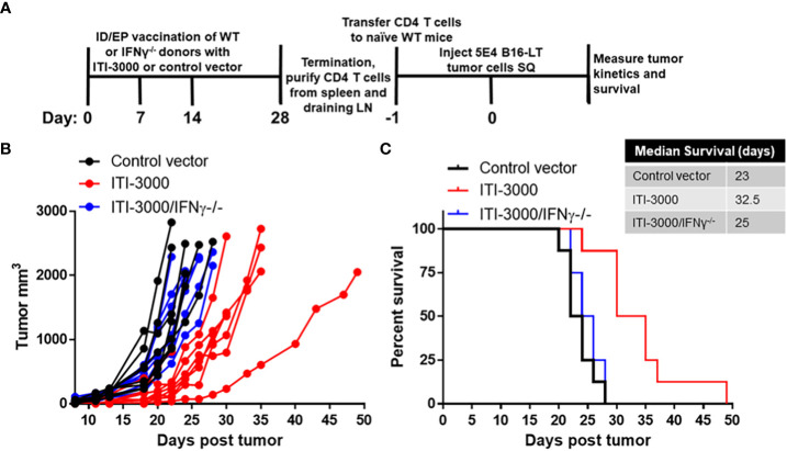
Methods: We designed a cancer vaccine that promotes potent, antigen-specific CD4 T cell responses to MCPyV-LT. To activate antigen-specific CD4 T cells in vivo, we utilized our nucleic acid platform, UNITE™ (UNiversal Intracellular Targeted Expression), which fuses a tumor-associated antigen with lysosomal-associated membrane protein 1 (LAMP1). This lysosomal targeting technology results in enhanced antigen presentation and potent antigen-specific T cell responses. LTS220A, encoding a mutated form of MCPyV-LT that diminishes its pro-oncogenic properties, was introduced into the UNITE™ platform.

Results: Vaccination with LTS220A-UNITE™ DNA vaccine (ITI-3000) induced antigen-specific CD4 T cell responses and a strong humoral response that were sufficient to delay tumor growth of a B16F10 melanoma line expressing LTS220A. This effect was dependent on the CD4 T cells' ability to produce IFNγ. Moreover, ITI-3000 induced a favorable tumor microenvironment (TME), including Th1-type cytokines and significantly enhanced numbers of CD4 and CD8 T cells as well as NK and NKT cells. Additionally, ITI-3000 synergized with an α-PD-1 immune checkpoint inhibitor to further slow tumor growth and enhance survival.

Conclusions: These findings strongly suggest that in pre-clinical studies, DNA vaccination with ITI-3000, using the UNITE™ platform, enhances CD4 T cell responses to MCPyV-LT that result in significant anti-tumor immune responses. These data support the initiation of a first-in-human (FIH) Phase 1 open-label study to evaluate the safety, tolerability, and immunogenicity of ITI-3000 in patients with polyomavirus-positive MCC (NCT05422781).

Keywords: Merkel cell carcinoma; Merkel cell polyomavirus; cancer vaccine; immunotherapy; tumor microenvironment.


Conflict of interest statement

TH is a co-founder and shareholder at Immunomic Therapeutics Inc. TH, CB, EL, and JH are employees of Immunomic Therapeutics Inc. The remaining authors declare that the research was conducted in the absence of any commercial or financial relationships that could be construed as a potential conflict of interest.

Figures

Figure 1
Figure 1
ITI-3000 slows tumor growth and enhances survival in both prophylactic and therapeutic settings. Experimental design for prophylactic vaccination shown in (A) C57BL/6 mice were vaccinated three times, weekly, with 40µg of ITI-3000 or control vector via intradermal injection followed by electroporation. Fourteen days following the final vaccination, 2E5 B16-LT tumor cells were injected subcutaneously in the right flank. Tumor kinetics (B) and survival (C) were measured over time. N=7. Representative of three separate experiments. Survival curve significance was calculated using a Log-rank (Mantel-Cox) test. Experimental design for therapeutic vaccination shown in (D) C57BL/6 mice were given 5E4 B16-LT tumor cells subcutaneously in the right flank. Starting on day 3 post tumor injection, mice were vaccinated four times, weekly, with 40µg of ITI-3000 or control vector via intradermal injection followed by electroporation. Tumor kinetics (E) and survival (F) were measured over time. N=26 control vector, N=22 ITI-3000, combined data from three experiments. Survival curve significance was calculated using a Log-rank (Mantel-Cox) test.
Figure 2
Figure 2
ITI-3000 induces antigen-specific CD4 T cells and anti-LT antibodies. Experimental design shown in (A) C57BL/6 mice were vaccinated four times, weekly, with 40µg of ITI-3000 or control vector via intradermal injection followed by electroporation. Mice were euthanized fourteen days following the final vaccination, and antigen-specific IFNγ peptide recall responses were evaluated in splenocytes by ELISpot (B). An overlapping peptide library spanning the large T antigen of MCPyV (five separate peptide pools) was used as stimulation at 2µg/ml. Media alone was used as a negative control. Data is represented as spot-forming units (SFU) per 1E6 splenocytes. Representative of three separate experiments. For intracellular cytokine staining (ICS), splenocytes were incubated for five hours in the presence of brefeldin A, monensin, and 2µg/ml LT peptides (peptide pool 2). Cells were then stained with fluorochrome-conjugated antibodies and analyzed using a CytoFLEX flow cytometer. Data is represented as percent cytokine-positive CD4 (C) and CD8 (D) effector/memory T cells (CD44+CD62Llo) N=8. Representative of three separate experiments. Statistical significance was calculated by two-way ANOVA. Antibody responses to different regions of the LT protein were evaluated by incubating serum with fluorescently-labeled beads loaded with GST-fusion proteins (the truncated large T antigen (tLT), or small T antigen (ST)). To confirm antibody reactivity to the unique region of LT (F), serum from ITI-3000-vaccinated mice (E) was pretreated with soluble GST-common T region before addition of T antigen-loaded beads. N=8. Representative of three separate experiments. Statistical significance was calculated by unpaired Student’s t-test. The "*" symbol represents a p-value of <0.05.
Figure 3
Figure 3
ITI-3000 induces immune cell infiltration into tumors. Mice were given 5E4 B16-LT tumor cells subcutaneously in the right flank. Starting on day 3 post tumor injection, mice were vaccinated twice, weekly, with 40µg of ITI-3000 or control vector via intradermal injection followed by electroporation. Three days following the second vaccination, mice were euthanized, and tumors were enzymatically digested and processed into a single cell suspension. The cellular composition of the tumor microenvironment, including numbers of CD45+ cells and CD3+ cells (A), numbers (B) and frequencies (C) of CD4+, CD8+, and CD4+Foxp3+CD25+ T cells, numbers of NK and NKT cells (D), and numbers and frequency of F4/80+ macrophages (E), was analyzed by flow cytometry. Data is represented as total number of cells per gram tumor tissue or as frequency of total CD45+ or CD4+ cells. N=8. Representative of two separate experiments. Statistical significance was calculated by unpaired Student’s t-test. The "*" symbol represents a p-value of <0.05. "NS" denotes a non-significant difference between groups.
Figure 4
Figure 4
ITI-3000 induces a systemic Th1-type cytokine profile with enhanced pro-inflammatory cytokines in the tumor microenvironment. Mice were given 5E4 B16-LT tumor cells subcutaneously in the right flank. Starting on day 3 post tumor injection, mice were vaccinated twice, weekly, with 40µg of ITI-3000 or control vector via intradermal injection followed by electroporation. Three days following the second vaccination, mice were euthanized, and serum and tumors were collected. Tumors were lysed and homogenized, and cytokine content of tumor extracts (A) and serum (B) was analyzed using the Meso Scale Discovery V-PLEX Proinflammatory Panel 1 Mouse Kit. N=10. Data is combined from two separate experiments. Statistical significance was calculated by unpaired Student’s t-test. The "*" symbol represents a p-value of <0.05.
Figure 5
Figure 5
ITI-3000 induces spatial immune T cell infiltration into tumors. Experimental design shown in (A). C57BL/6 mice were given 5E4 B16-LT tumor cells subcutaneously in the right flank. Starting on day 3 post tumor injection, mice were vaccinated twice, weekly, with 40µg of ITI-3000 or control vector via intradermal injection followed by electroporation. Tumors were collected and formalin fixed on day 15 post tumor injection, followed by a sucrose gradient, paraffin embedding, and sectioning. The cellular composition of the tumor microenvironment on FFPE slides was determined by multiplex-IHC including percent of CD3+ (green), CD8+ (magenta) (B), and CD4+ (red) (C) cells. One representative section (displayed both as whole tumor section (right) and magnified area of interest (left)) of each group is shown in (D) The total cell population was determined by nuclear DAPI staining (blue). Sections were analyzed by AI gating in the Biodock platform. N=5. Statistical significance was calculated by unpaired Student’s t-test. The "*" symbol represents a p-value of <0.05.
Figure 6
Figure 6
ITI-3000-mediated tumor control is dependent on IFNγ production by antigen-specific CD4 T cells. Experimental design shown in (A). C57BL/6 donor mice (WT or IFNγ-/-) were vaccinated three times, weekly, with 40µg of ITI-3000 or control vector via intradermal injection followed by electroporation. Mice were euthanized fourteen days following the final vaccination, and total CD4 T cells were purified by magnetic bead negative selection from spleens and draining cervical LN, pooled within groups. 10E6 CD4 T cells, either WT or IFNγ-/-, were transferred IV into naïve WT C56BL/6 recipient mice. One day following CD4 T cell transfer, 5E4 B16-LT tumor cells were injected subcutaneously in the right flank. Tumor kinetics (B) and survival (C) were measured over time. N=8. Representative of two separate experiments. Survival curve significance was calculated using a Log-rank (Mantel-Cox) test corrected for multiple comparisons. Control vector vs ITI-3000 p = 0.0003, ITI-3000 vs ITI-3000/IFNγ-/- p = 0.0007, Control vector vs ITI-3000/IFNγ-/- p = 0.29.
Figure 7
Figure 7
ITI-3000 synergizes with α-PD-1 checkpoint blockade therapy to further slow tumor growth and enhance survival. Experimental design shown in (A). C57BL/6 mice were given 5E4 B16-LT tumor cells subcutaneously in the right flank. Starting on day 3 post tumor injection, mice were vaccinated four times, weekly, with 40µg of ITI-3000 or control vector via intradermal injection followed by electroporation. On the same dates, mice were also given 200µg of either α-PD-1 monoclonal antibody or of an isotype control. Tumor kinetics (B) and survival (C) were measured over time. N=14. Combined data from two experiments. Survival curve significance was calculated using a Log-rank (Mantel-Cox) test corrected for multiple comparisons. Control vector/isotype vs ITI-3000/isotype p = 0.0153, Control vector/isotype vs ITI-3000/α-PD-1 p = <0.0001, ITI-3000/isotype vs ITI-3000/α-PD-1 p = 0.0267. The "*" symbol represents a p-value of <0.05.

Similar articles

Cited by

References

    1. Becker JC, Stang A, DeCaprio JA, Cerroni L, Lebbé C, Veness M, et al. . Merkel cell carcinoma. Nat Rev Dis Primers (2017) 3:17077. doi: 10.1038/nrdp.2017.77 - DOI - PMC - PubMed
    1. Chang Y, Moore PS. Merkel cell carcinoma: a virus-induced human cancer. Annu Rev Pathol (2012) 7:123–44. doi: 10.1146/annurev-pathol-011110-130227 - DOI - PMC - PubMed
    1. Paulson KG, Bhatia S. Advances in immunotherapy for metastatic Merkel cell carcinoma: a clinician’s guide. J Natl Compr Cancer Netw: JNCCN (2018) 16(6):782–90. doi: 10.6004/jnccn.2018.7049 - DOI - PubMed
    1. Goh G, Walradt T, Markarov V, Blom A, Riaz N, Doumani R, et al. . Mutational landscape of MCPyV-positive and MCPyV-negative Merkel cell carcinomas with implications for immunotherapy. Oncotarget (2016) 7(3):3403–15. doi: 10.18632/oncotarget.6494 - DOI - PMC - PubMed
    1. Agelli M, Clegg LX. Epidemiology of primary Merkel cell carcinoma in the United States. J Am Acad Dermatol (2003) 49(5):832–41. doi: 10.1016/S0190-9622(03)02108-X - DOI - PubMed

Publication types

Substances

Associated data