

Nanostructured N/S doped carbon dots/mesoporous silica nanoparticles and PVA composite hydrogel fabrication for anti-microbial and anti-biofilm application
- PMID: 37711848
- PMCID: PMC10498006
- DOI: 10.1016/j.ijpx.2023.100209
Nanostructured N/S doped carbon dots/mesoporous silica nanoparticles and PVA composite hydrogel fabrication for anti-microbial and anti-biofilm application
Abstract
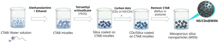
Regarding the convergence of the worldwide epidemic, the appearance of bacterial infection has occasioned in a melodramatic upsurge in bacterial pathogens with confrontation against one or numerous antibiotics. The implementation of engineered nanostructured particles as a delivery vehicle for antimicrobial agent is one promising approach that could theoretically battle the setbacks mentioned. Among all nanoparticles, silica nanoparticles have been found to provide functional features that are advantageous for combatting bacterial contagion. Apart from that, carbon dots, a zero-dimension nanomaterial, have recently exhibited their photo-responsive property to generate reactive oxygen species facilitating to enhance microorganism suppression and inactivation ability. In this study, potentials of core/shell mesoporous silica nanostructures (MSN) in conjugation with carbon dots (CDs) toward antimicrobial activity against Staphylococcus aureus, Pseudomonas aeruginosa and Escherichia coli have been investigated. Nitrogen and sulfur doped CDs (NS/CDs) conjugated with MSN which were cost effective nanoparticles exhibited much superior antimicrobial activity for 4 times as much as silver nanoparticles against all bacteria tested. Among all nanoparticles tested, 0.40 M NS/CDs@MSN showed the greatest minimal biofilm inhibitory at very low concentration (< 0.125 mg mL-1), followed by 0.20 M NS/CDs@MSN (0.5 mg mL-1), CD@MSN (25 mg mL-1), and MSN (50 mg mL-1), respectively. Immobilization of NS/CDs@MSN in polyvinyl alcohol (PVA) hydrogel was performed and its effect on antimicrobial activity, biofilm controlling efficiency, and cytotoxicity toward fibroblast (NIH/3 T3 and L-929) cells was additionally studied for further biomedical applications. The results demonstrated that 0.40 M NS/CDs-MSN@PVA hydrogel exhibited the highest inhibitory effect on S. aureus > P. aeruginosa > E. coli. In addition, MTT assay revealed some degree of toxicity of 0.40 M NS/CDs-MSN@PVA hydrogel against L-929 cells by a slight reduction of cell viability from 100% to 81.6% when incubated in the extract from 0.40 M NS/CDs-MSN@PVA hydrogel, while no toxicity of the same hydrogel extract was detected toward NIH/3 T3 cells.
Keywords: Antimicrobial activity; Carbon dots; Cytotoxicity test; Freeze-thaw technique; Health and well-being; MTT assay; Mesoporous silica nanostructures; Polyvinyl alcohol hydrogel.
© 2023 Published by Elsevier B.V.
Conflict of interest statement
The authors declare that they have no known competing financial interests or personal relationships that could have appeared to influence the work reported in this paper.
Figures


References
-
- Abbasi M., Gholizadeh R., Kasaee S.R., Vaez A., Chelliapan S., Fadhil Al-Qaim F., Deyab I.F., Shafiee M., Zareshahrabadi Z., Amani A.M., Mosleh-Shirazi S., Kamyab H. An intriguing approach toward antibacterial activity of green synthesized Rutin-templated mesoporous silica nanoparticles decorated with nanosilver. Sci. Rep. 2023;13:5987. - PMC - PubMed
-
- Auriemma F., De Rosa C., Ricciardi R., Lo Celso F., Triolo R., Pipich V. Time-resolving analysis of cryotropic gelation of water/poly(vinyl alcohol) solutions via small-angle neutron scattering. J. Phys. Chem. B. 2008;112:816–823. - PubMed
-
- Baig M.M., Zulfiqar S., Yousuf M.A., Shakir I., Aboud M.F.A., Warsi M.F. DyxMnFe2-xO4 nanoparticles decorated over mesoporous silica for environmental remediation applications. J. Hazard. Mater. 2021;402 - PubMed
-
- Bernardos A., Piacenza E., Sancenón F., Hamidi M., Maleki A., Turner R.J., Martínez-Máñez R. Mesoporous silica-based materials with bactericidal properties. Small. 2019;15:1900669. - PubMed
LinkOut - more resources
Full Text Sources
Research Materials
Miscellaneous
