. 2023 Aug 30:14:1234804.
doi: 10.3389/fgene.2023.1234804. eCollection 2023.

Clinical and molecular delineation of classical-like Ehlers-Danlos syndrome through a comprehensive next-generation sequencing-based screening system

Affiliations

Clinical and molecular delineation of classical-like Ehlers-Danlos syndrome through a comprehensive next-generation sequencing-based screening system

Tomomi Yamaguchi et al. Front Genet. .

Abstract

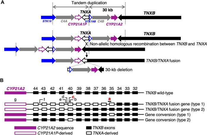
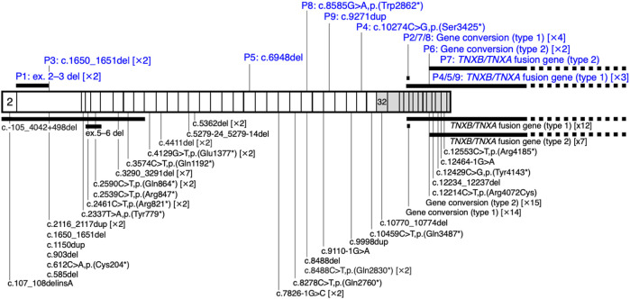
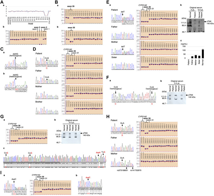
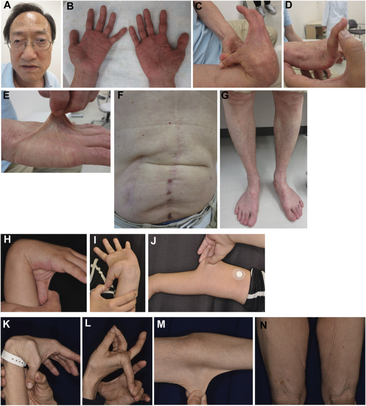
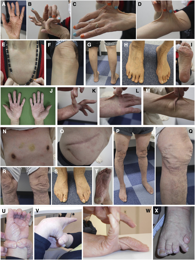
Classical-like Ehlers-Danlos syndrome (clEDS) is an autosomal recessive disorder caused by complete absence of tenascin-X resulting from biallelic variation in TNXB. Thus far, 50 patients from 43 families with biallelic TNXB variants have been identified. Accurate detection of TNXB variants is challenging because of the presence of the pseudogene TNXA, which can undergo non-allelic homologous recombination. Therefore, we designed a genetic screening system that is performed using similar operations to other next-generation sequencing (NGS) panel analyses and can be applied to accurately detect TNXB variants and the recombination of TNXA-derived sequences into TNXB. Using this system, we identified biallelic TNXB variants in nine unrelated clEDS patients. TNXA-derived variations were found in >75% of the current cohort, comparable to previous reports. The current cohort generally exhibited similar clinical features to patients in previous reports, but had a higher frequency of gastrointestinal complications (e.g., perforation, diverticulitis, gastrointestinal bleeding, intestinal obstruction, rectal/anal prolapse, and gallstones). This report is the first to apply an NGS-based screening for TNXB variants and represents the third largest cohort of clEDS, highlighting the importance of increasing awareness of the risk of gastrointestinal complications.

Keywords: Ehlers-Danlos syndrome; TNXB; classical-like; connective tissue disorder; tenascin-X.


Conflict of interest statement

TY, TF, YT, and TK are members of the endowed chair named “Division of Clinical Sequencing, Shinshu University School of Medicine”, which is sponsored by BML, Inc. and Life Technologies Japan Ltd., a subsidiary of Thermo Fisher Scientific Inc. The remaining author declares that the research was conducted in the absence of any commercial or financial relationships that could be construed as a potential conflict of interest.

Figures

FIGURE 1
FIGURE 1
Schematic diagram of TNXB/TNXA fusion genes. (A) TNXB/TNXA fusion genes resulting from non-allelic homologous recombination between the TNXA pseudogene and TNXB, resulting in 30-kb deletions. (B) TNXB/TNXA fusion genes (type 1 and type 2) and gene conversions (type 1 and type 2). Seven indices of TNXA-derived variations are shown: a, 120-bp deletion (c.11435_11524 + 30del) in exon 35–intron 35; b, c.12150C>G,p.(Arg4050 = ) in exon 40; c, c.12174C>G,p.(Cys4058Trp) in exon 40; d, c.12204 + 39dup in intron 40; e, c.12204 + 43T>G in intron 40; f, c.12628-52A>G in intron 43; g, CYP21A2 deletion. Only a 120-bp deletion in exon 35–intron 35 and c.12174C>G in exon 40 shown in red have been reported to be pathogenic (Demirdas et al., 2017).
FIGURE 2
FIGURE 2
Schematic representation of the distribution of TNXB variants in a cohort of nine patients with clEDS. Illustration of TNXB variants based on NM_019105.6 of the NCBI reference sequence database (https://www.ncbi.nlm.nih.gov/RefSeq/). Exons 32–44 are highlighted with gray boxes. Variants found in the current study of patients (P)1–9 are shown in blue, above the mRNA transcript. Previously reported variants (Burch et al., 1997; Schalkwijk et al., 2001; Voermans et al., 2009; Hendriks et al., 2012; Pénisson-Besnier et al., 2013; Sakiyama et al., 2015; Chen et al., 2016; Demirdas et al., 2017; Micale et al., 2019; Rymen et al., 2019; Brisset et al., 2020; Green et al., 2020; Colman et al., 2021; Watanabe et al., 2021; Al-Harbi et al., 2022; Santoreneos et al., 2022) are shown below the mRNA transcript.
FIGURE 3
FIGURE 3
Molecular investigation of Patients 1–9. (A) Patient 1: homozygosity for exons 2–3 deletion detected by the CNV visualization method for an amplification-based NGS data (a) and validated by MLPA (b). (B) Patient 2: homozygosity for a gene conversion characterized by TNXA-derived variation with validation of the exon 35 deletion by MLPA in the patient (a) and his son (b). (C) Patient 3: homozygosity for a frameshift variant c.1650_1651del,p.(Glu552Argfs*41) validated by Sanger sequencing in the patient (a) and his daughter (b). (D) Patient 4: compound heterozygosity for a nonsense variant c.10274C>G,p.(Ser3425*) and a TNXB/TNXA fusion gene characterized by TNXA-derived variation with validation of the nonsense variant by Sanger sequencing (left) and the TNXB exon 35 deletion and CYP21A2 deletion by MLPA (right) in the patient and her family. (E) Patient 5: compound heterozygosity for a frameshift variant c.6948del,p.(Asp2317Thrfs*53) and a TNXB/TNXA fusion gene characterized by TNXA-derived variation with validation of the frameshift variant by Sanger sequencing (left) and the TNXB exon 35 deletion and CYP21A2 deletion by MLPA (right) in the patient and her family (a). Western blot analysis of sTNX in the patient (b) and quantification of sTNX in the patient and her family using nano-LC/MS/MS (c). (F) Patient 6: homozygosity for a gene conversion characterized by TNXA-derived variation with validation by Sanger sequencing (a) and triplicate Western blot analysis of sTNX in the patient (b). (G) Patient 7: compound heterozygosity for a gene conversion and a TNXB/TNXA fusion gene characterized by TNXA-derived variation with validation of the TNXB exon 35 deletion and CYP21A2 deletion by MLPA (a). Triplicate Western blot analysis of sTNX (b). Sanger sequencing of the region around exon 40 in the normal exon 35 allele (c), with the upper row showing the TNXB sequence and the lower row showing the TNXA sequence. TNXB exon 40 is highlighted with a gray box. (H) Patient 8: compound heterozygosity for a nonsense variant c.8585G>A,p.(Trp2862*) and a gene conversion characterized by TNXA-derived variation with validation of the nonsense variant by Sanger sequencing (left) and the TNXB exon 35 deletion by MLPA (right) in the patient and her parents. (I) Patient 9: compound heterozygosity for a frameshift variant c.9271dup,p.(Gln3091Profs*31) and a TNXB/TNXA fusion gene characterized by TNXA-derived variation with validation of the frameshift variant by Sanger sequencing (left) and the TNXB exon 35 deletion and CYP21A2 deletion by MLPA (right) (a). Sanger sequencing of the frameshift variant c.9271dup,p.(Gln3091Profs*31) in the normal exon 35 allele (b), with the upper row showing the TNXB sequence and the lower row showing the TNXA sequence. TNXB exon 27 is highlighted with a gray box.
FIGURE 4
FIGURE 4
Clinical photographs of Patients 2, 4, and 5. (A–G) Patient 2 at age 65 years, showing jaw protrusion (A), hands with thick fingers and wrinkled palms (B), hypermobile finger joints (C,D), hyperextensible skin (E), atrophic scars at surgical sutures (F), and valgus/flat feet (G). (H–J) Patient 4 at age 27 years, showing finger joint hypermobility (H,I) and skin hyperextensiblity (J). (K–N) Patient 5 at age 47 years, showing marked finger joint hypermobility (K,L) and skin hyperextensibility (M) and redundancy (N).
FIGURE 5
FIGURE 5
Clinical photographs of Patients 6, 7, and 9. (A–I) Patient 6 at age 59 years, showing wrinkled palms (A), hypermobile finger joints (B), skin hyperextensibility, translucency, and bruisability (C–G), mildly atrophic thighs (G), pes planus (H), and moderate calluses on the soles (I). (J–T) Patient 7 at age 61 years, showing wrinkled palms (J), mild joint hypermobility with a Beighton score of 4/9 (K), hyperextensible, bruisable, thin, velvety, and translucent skin (L–N,R), without fragility or atrophic scars (O), mild pectus excavatum (N), atrophic thighs (P), multiple varices at the knees (Q), pes planus (S), and severe calluses on the soles (T). (U–X) Patient 9 at age 69 years, showing brachydactyly with excessive skin (U,W) and toe deformities (X), but no generalized joint hypermobility (V,W).

Similar articles

Cited by

References

    1. Al-Harbi T. M., Al-Rammah H., Al-Zahrani N., Liu Y., Sleiman P. M. A., Dridi W., et al. (2022). Rare neurological manifestations in a Saudi Arabian patient with Ehlers–Danlos syndrome and a novel homozygous variant in the TNXB gene. Am. J. Med. Genet. A 188 (2), 618–623. 10.1002/ajmg.a.62539 - DOI - PubMed
    1. Brady A. F., Demirdas S., Fournel-Gigleux S., Ghali N., Giunta C., Kapferer-Seebacher I., et al. (2017). The Ehlers–Danlos syndromes, rare types. Am. J. Med. Genet. C Semin. Med. Genet. 175, 70–115. 10.1002/ajmg.c.31550 - DOI - PubMed
    1. Brisset M., Metay C., Carlier R. Y., Badosa C., Marques C., Schalkwijk J., et al. (2020). Biallelic mutations in Tenascin-X cause classical-like Ehlers–Danlos syndrome with slowly progressive muscular weakness. Neuromuscul. Disord. 30 (10), 833–838. 10.1016/j.nmd.2020.09.002 - DOI - PubMed
    1. Burch G. H., Gong Y., Liu W., Dettman R. W., Curry C. J., Smith L., et al. (1997). Tenascin-X deficiency is associated with Ehlers–Danlos syndrome. Nat. Genet. 17, 104–108. 10.1038/ng0997-104 - DOI - PubMed
    1. Chen W., Perritt A. F., Morissette R., Dreiling J. L., Bohn M. F., Mallappa A., et al. (2016). Ehlers–Danlos syndrome caused by biallelic TNXB variants in patients with congenital adrenal hyperplasia. Hum. Mutat. 37 (9), 893–897. 10.1002/humu.23028 - DOI - PMC - PubMed