

Clinical and genetic features of Koreans with retinitis pigmentosa associated with mutations in rhodopsin
- PMID: 37712069
- PMCID: PMC10497939
- DOI: 10.3389/fgene.2023.1240067
Clinical and genetic features of Koreans with retinitis pigmentosa associated with mutations in rhodopsin
Abstract
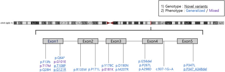
Purpose: To investigate the clinical features, natural course, and genetic characteristics of Koreans with rhodopsin-associated retinitis pigmentosa (RHO-associated RP). Design: We conducted a retrospective, multicenter, observational cohort study. Participants: We reviewed the medical records of 42 patients with RHO-associated RP of 36 families who visited 4 hospitals in Korea. Methods: Patients with molecular confirmation of pathogenic variants of the RHO gene were included. The patients were divided into two subgroups: the generalized and sector RP groups. A central visual field of the better-seeing eye of <10° or a best-corrected visual acuity of the better-seeing eye <20/40 indicated the progression to late-stage RP. Results: The mean age at which symptoms first appeared was 26.3 ± 17.9 years (range: 8-78 years), and the mean follow-up period was 80.9 ± 68.7 months (range: 6-268 months). At the last follow-up visit, the generalized RP group showed a significantly higher rate of visual field impairment progression to late-stage RP than that of the sector RP group (22 of 35 [62.9%] vs. 0 of 7 [0.0%], p = 0.003). No cases in the sector RP group progressed to generalized RP. Best-corrected visual acuity deterioration to late-stage RP was observed only in the generalized RP group (13 of 35 patients; 37.1%), whereas no deterioration was observed in the sector RP group. We identified 16 known and three novel RHO mutations, including two missense mutations (p.T108P and p.G121R) and one deletion mutation (p.P347_A348del). The pathogenic variants were most frequently detected in exon 1 (14 of 36 [38.9%]). The most common pathogenic variants were p.P347L and T17M (5 of 36 [13.9%] families). Among 42 patients of 36 families, 35 patients of 29 families (80.6%) presented with the generalized RP phenotype, and seven patients of seven families (19.4%) presented with the sector RP phenotype. Three variants (p.T17M, p.G101E, and p.E181K) presented with both the generalized and sector RP phenotypes. Conclusion: This multicenter cohort study provided information on the clinical and genetic features of RHO-associated RP in Koreans. It is clinically important to expand the genetic spectrum and understand genotype-phenotype correlations to ultimately facilitate the development of gene therapy.
Keywords: Koreans; generalized retinitis pigmentosa; retinitis pigmentosa; rhodopsin; sector retinitis pigmentosa.
Copyright © 2023 Jung, Kwak, Joo, Lee, Park, Kim, Lee, Byeon, Lee, Han, Lee, Yoon and Woo.
Conflict of interest statement
The authors declare that the research was conducted in the absence of any commercial or financial relationships that could be construed as a potential conflict of interest.
Figures
Similar articles
-
Genotypes and clinical features of RHO-associated retinitis pigmentosa in a Japanese population.Jpn J Ophthalmol. 2024 Jan;68(1):1-11. doi: 10.1007/s10384-023-01036-0. Epub 2023 Dec 9. Jpn J Ophthalmol. 2024. PMID: 38070066
-
Spectrum of rhodopsin mutations in Korean patients with retinitis pigmentosa.Mol Vis. 2011;17:844-53. Epub 2011 Apr 1. Mol Vis. 2011. PMID: 21677794 Free PMC article.
-
Beyond Sector Retinitis Pigmentosa: Expanding the Phenotype and Natural History of the Rhodopsin Gene Codon 106 Mutation (Gly-to-Arg) in Autosomal Dominant Retinitis Pigmentosa.Genes (Basel). 2021 Nov 23;12(12):1853. doi: 10.3390/genes12121853. Genes (Basel). 2021. PMID: 34946802 Free PMC article.
-
Sector retinitis pigmentosa: Report of ten cases and a review of the literature.Mol Vis. 2019 Dec 30;25:869-889. eCollection 2019. Mol Vis. 2019. PMID: 31908405 Free PMC article. Review.
-
Vitamin A and fish oils for preventing the progression of retinitis pigmentosa.Cochrane Database Syst Rev. 2020 Jun 18;6(6):CD008428. doi: 10.1002/14651858.CD008428.pub3. Cochrane Database Syst Rev. 2020. PMID: 32573764 Free PMC article.
Cited by
-
RHO Variants and Autosomal Dominant Retinitis Pigmentosa: Insights from the Italian Genetic Landscape.Genes (Basel). 2024 Sep 2;15(9):1158. doi: 10.3390/genes15091158. Genes (Basel). 2024. PMID: 39336749 Free PMC article.
-
Linking Protein Stability to Pathogenicity: Predicting Clinical Significance of Single-Missense Mutations in Ocular Proteins Using Machine Learning.Int J Mol Sci. 2024 Oct 30;25(21):11649. doi: 10.3390/ijms252111649. Int J Mol Sci. 2024. PMID: 39519200 Free PMC article.
References
-
- Ballios B. G., Place E. M., Martinez-Velazquez L., Pierce E. A., Comander J. I., Huckfeldt R. M. (2021). Beyond sector retinitis pigmentosa: expanding the phenotype and natural history of the rhodopsin gene codon 106 mutation (Gly-to-Arg) in autosomal dominant retinitis pigmentosa. Genes (Basel) 12, 1853. 10.3390/genes12121853 - DOI - PMC - PubMed
LinkOut - more resources
Full Text Sources