. 2023 Sep;13(9):e1410.
doi: 10.1002/ctm2.1410.

Long noncoding RNA Regulating ImMune Escape regulates mixed lineage leukaemia protein-1-H3K4me3-mediated immune escape in oesophageal squamous cell carcinoma

Affiliations

Long noncoding RNA Regulating ImMune Escape regulates mixed lineage leukaemia protein-1-H3K4me3-mediated immune escape in oesophageal squamous cell carcinoma

Jia Liu et al. Clin Transl Med. 2023 Sep.

Abstract

Background: Predictive biomarkers for oesophageal squamous cell carcinoma (ESCC) immunotherapy are lacking, and immunotherapy resistance remains to be addressed. The role of long noncoding RNA (lncRNA) in ESCC immune escape and immunotherapy resistance remains to be elucidated.

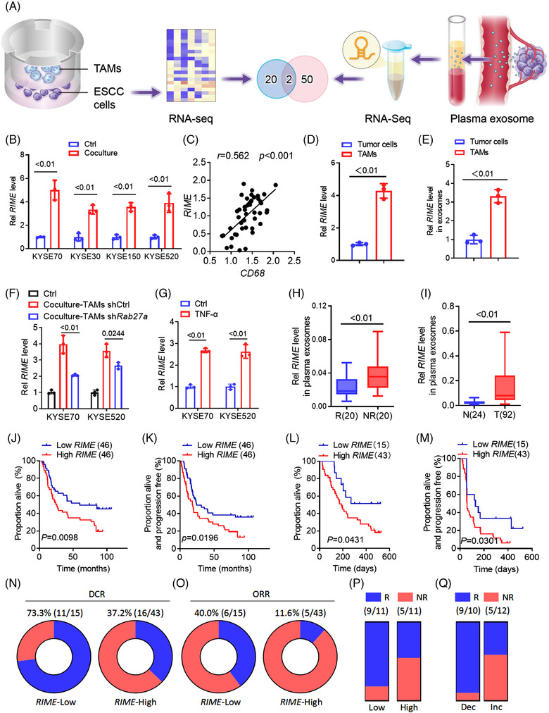
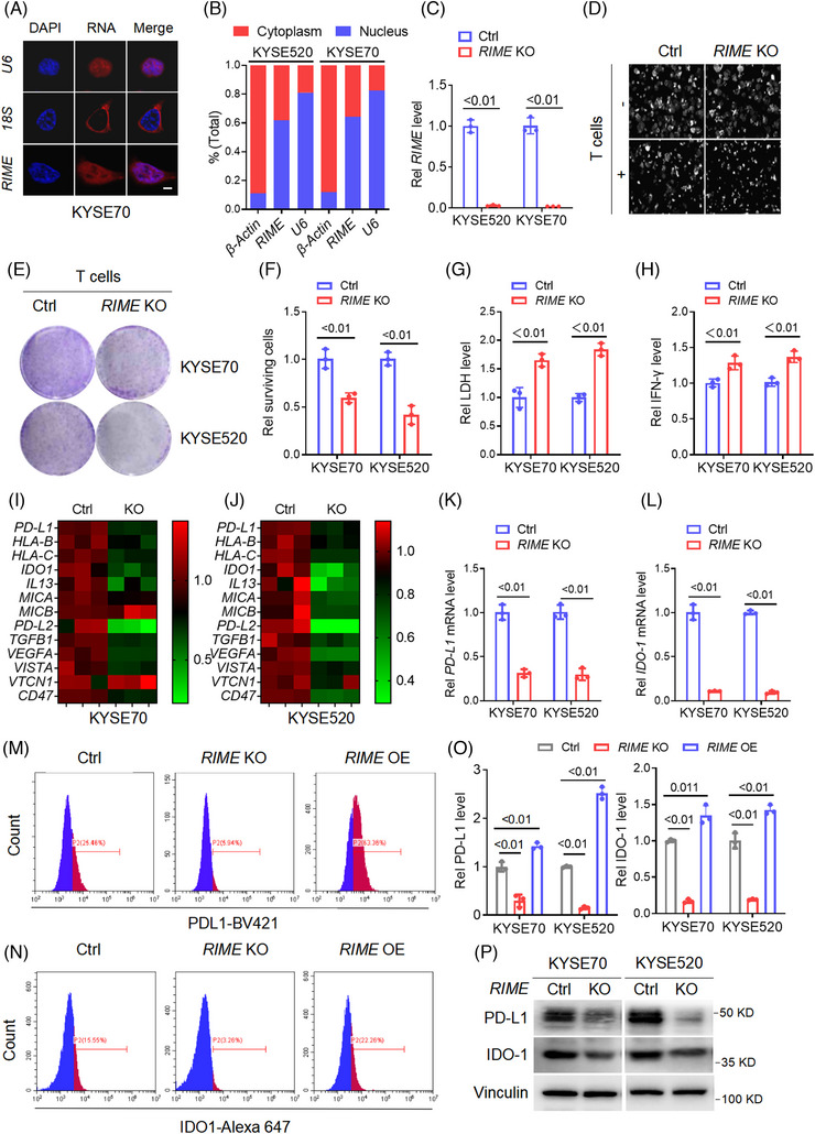
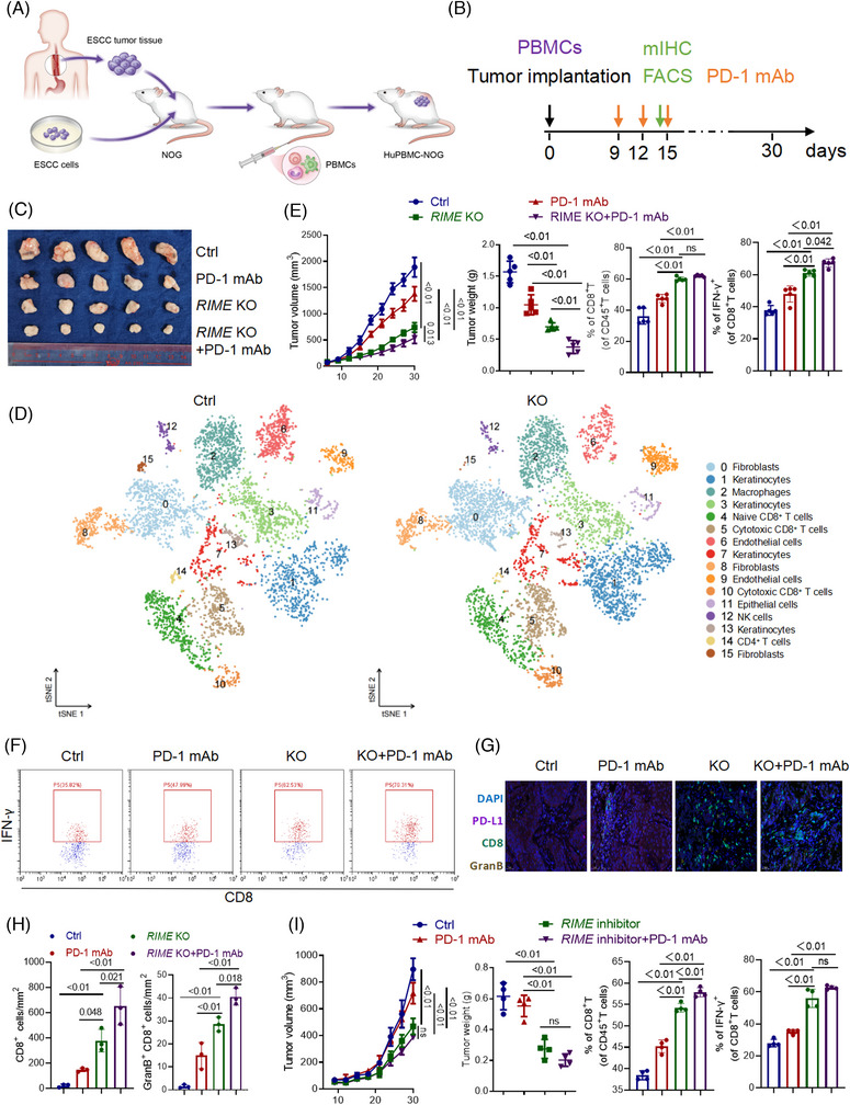
Methods: The tumour-associated macrophage-upregulated lncRNAs and the exosomal lncRNAs highly expressed in ESCC immunotherapy nonresponders were identified by lncRNA sequencing and polymerase chain reaction assays. CRISPR-Cas9 was used to explore the functional roles of the lncRNA. RNA pull-down, MS2-tagged RNA affinity purification (MS2-TRAP) and RNA-binding protein immunoprecipitation (RIP) were performed to identify lncRNA-associated proteins and related mechanisms. In vivo, the humanized PBMC (hu-PBMC) mouse model was established to assess the therapeutic responses of specific lncRNA inhibitors and their combination with programmed cell death protein 1 (PD-1) monoclonal antibody (mAb). Single-cell sequencing, flow cytometry, and multiplex fluorescent immunohistochemistry were used to analyze immune cells infiltrating the tumour microenvironment.

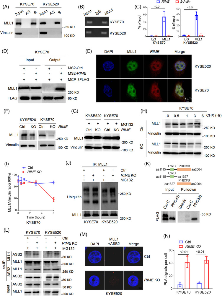
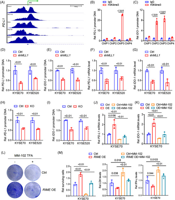
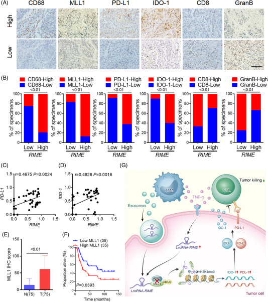
Results: We identified a lncRNA that is involved in tumour immune evasion and immunotherapy resistance. High LINC02096 (RIME) expression in plasma exosomes correlates with a reduced response to PD-1 mAb treatment and poor prognosis. Mechanistically, RIME binds to mixed lineage leukaemia protein-1 (MLL1) and prevents ankyrin repeat and SOCS box containing 2 (ASB2)-mediated MLL1 ubiquitination, improving the stability of MLL1. RIME-MLL1 increases H3K4me3 levels in the promoter regions of programmed death-ligand 1 (PD-L1) and indoleamine 2,3-dioxygenase 1 (IDO-1), constitutively increasing the expression of PD-L1/IDO-1 in tumour cells and inhibiting CD8+ T cells infiltration and activation. RIME depletion in huPBMC-NOG mice significantly represses tumour development and improves the effectiveness of PD-1 mAb treatment by activating T-cell-mediated antitumour immunity.

Conclusions: This study reveals that the RIME-MLL1-H3K4me3 axis plays a critical role in tumour immunosuppression. Moreover, RIME appears to be a potential prognostic biomarker for immunotherapy and developing drugs that target RIME may be a new therapeutic strategy that overcomes immunotherapy resistance and benefits patients with ESCC.

Keywords: esophageal squamous cell carcinoma; immune escape; immunotherapy; lncRNA; mixed lineage leukaemia protein-1 (MLL1).


Conflict of interest statement

The authors declare no conflict of interest.

Figures

FIGURE 1
FIGURE 1
LncRNA RIME correlates with immunotherapy outcomes. (A) Illustration of tumour‐associated macrophage (TAM)‐upregulated long noncoding RNAs (lncRNAs) and plasma exosomal lncRNAs upregulated in oesophageal squamous cell carcinoma (ESCC) immunotherapy nonresponders. (B) quantitative real‐time polymerase chain reaction (qRT‒PCR) analysis showed that RIME expression was upregulated in ESCC cells cocultured with TAMs from ESCC patients. Ctrl, control. (C) qRT‒PCR and Pearson correlation analysis showed that RIME and CD68 expression in ESCC tumour tissues was positively correlated. (D) qRT‐PCR analysis showed that RIME was more highly expressed in TAMs than in ESCC tumour cells. (E) qRT‒PCR analysis of RIME expression in exosomes isolated from ESCC cells and TAMs, which showed that RIME was more highly expressed in exosomes from TAMs. (F) qRT‒PCR analysis showed that RIME expression was only partially suppressed in ESCC cells co‐cultured with Rab27a‐depleted TAMs. (G) qRT‒PCR analysis showed that TNF‐α stimulation upregulated RIME expression in ESCC cells. (H) qRT‒PCR analysis showed that RIME expression was increased in the plasma exosomes of ESCC PD‐1 mAb non‐responders compared to responders. R, responders. NR, non‐responders. (I) qRT‒PCR analysis showed that RIME expression was increased in the plasma exosomes of ESCC patients compared to healthy donors. T, ESCC patients. N, healthy donors. (J, K) OS and PFS analysis showed that high RIME expression in plasma correlated with poor OS and PFS of ESCC patients. (L, M) OS and PFS analysis showed that high RIME expression in plasma shortened OS and PFS of ESCC patients treated with PD‐1 mAb. (N, O) The disease control rate (DCR) and objective response rate (ORR) of PD‐1 mAb treatment was lower in the RIME high expression group. (P) The response rate of PD‐1 mAb plus chemotherapy (TP) in the plasma RIME high expression group was lower than RIME low expression group. (Q) The response rate of PD‐1 mAb plus chemotherapy (TP) in the plasma RIME increased (Inc) or decreased (Dec) group. Plasma RIME levels were examined before and after PD‐1 mAb plus chemotherapy (TP) treatment. qRT‐PCR analysis showed that the response rate was lower in the RIME‐increased group.
FIGURE 2
FIGURE 2
RIME reduces sensitivity to the cytotoxicity of CD8+ T cells by regulating immune checkpoint molecules. (A) Fluorescence in situ hybridization (FISH) assays showing the subcellular localization of RIME was both in the nucleus and cytoplasm of oesophageal squamous cell carcinoma (ESCC) cells. Scale bar, 2 μm. (B) Quantitative real‐time polymerase chain reaction (qRT‒PCR) analysis of RIME expression in the cytoplasmic and nuclear fractions of ESCC cells, which showed that RIME was localized both in the nucleus and cytoplasm. (C) qRT‒PCR analysis showed that RIME expression was significantly inhibited in RIME CRISPR KO cells. (D) Real‐time cell analyser was used to analyse the viability of ESCC cells alone or cocultured with human CD8+ T cells. RIME KO had a slight effect on ESCC cell viability but significantly enhanced the cytotoxicity of CD8+ T cells. (E, F) The cytotoxicity of CD8+ T cells to ESCC cells was determined by crystal violet staining, which showed that RIME KO rendered ESCC cells more sensitive to cytotoxic CD8+ T cells. (G) The LDH levels released from damaged cells were determined to quantify the cytotoxicity of CD8+ T cells. The LDH levels were significantly increased in the RIME KO group. (H) ELISA assays showed that the IFN‐γ levels in the coculture medium were significantly increased in the RIME KO group. (I, J) qRT‒PCR analysis showed that RIME KO significantly decreased multiple immune‐related genes in ESCC cells. (K, L) qRT‒PCR analysis showed that RIME KO resulted in a remarkable decrease of PD‐L1 and IDO‐1 expression in ESCC cells. (M–O) Flow cytometry and statistical analysis showed that RIME KO significantly decreased PD‐L1 and IDO‐1 expression, while RIME overexpression increased PD‐L1 and IDO‐1 expression. (P) Immunoblot analysis showed that RIME KO significantly decreased PD‐L1 and IDO‐1 protein levels in ESCC cells.
FIGURE 3
FIGURE 3
RIME binds and stabilizes MLL1. (A) RNA pulldown assays and immunoblot analysis showed that RIME specifically bound to MLL1. S, sense; AS, antisense. (B, C) RNA immunoprecipitation assays and quantitative real‐time polymerase chain reaction (qRT‒PCR) analysis verified the interaction of RIME and MLL1. (D) MS2‐tagged RNA affinity purification assays and immunoblot analysis verified the interaction of RIME and MLL1 in vivo. (E) Fluorescent in situ hybridization and immunofluorescence analysis showed that RIME and MLL1 colocalized mainly in the nucleus. (F) Immunoblot analysis showed that RIME KO remarkably reduced the MLL1 protein levels in oesophageal squamous cell carcinoma (ESCC) cells. (G) Immunoblot analysis showed that the decreased MLL1 levels in the RIME KO cells were recovered by the proteasomal inhibitor MG‐132 (10 μM, 12 h). (H‐I) Immunoblot analysis and quantification of MLL1 protein levels in KYSE70 cells treated with CHX (100 μg/ml) for the indicated times. As shown, RIME KO significantly shortened the half‐life of MLL1 in ESCC cells. (J) Immunoprecipitation and immunoblot analysis showed that RIME KO increased the ubiquitin levels of MLL1. (K) Schematic diagram of the MLL1 domains (top) and RNA pull‐down analysis of RIME‐associated FLAG‐tagged MLL1 domains (bottom). It showed that the PHD/Bromo fragment of MLL1 is required for the interaction of RIME and MLL1. (L) Co‐IP and immunoblot analysis showed that RIME KO significantly increased the MLL1/ASB2 interaction in ESCC cells. (M, N) Duolink proximity ligation assay and statistical analysis showed that RIME KO significantly increased the MLL1/ASB2 interaction in ESCC cells. Scale bar, 2 μm.
FIGURE 4
FIGURE 4
RIME regulates MLL1‐H3K4me3‐mediated PD‐L1/IDO‐1 expression. (A) ChIP‐seq analysis showed that H3K4me3 levels were enriched in the PD‐L1 promoter regions. The ChIP‐seq data was obtained from ENCODE database. (B‐C) ChIP assays indicated that H3K4me3 marks were enriched in the PD‐L1 promoter region around −1000 to 0 bp (ChIP1 to ChIP4 are primers designed for each fragment, representing −3000 to −2000 bp, −2000 to −1000 bp, −1000 to 0 bp, and 0 to +1000 bp, respectively), and in the IDO‐1 promoter region around −1500 to −500 bp (ChIP1 to ChIP4 represented −2000 to 0 bp). (D‐E) ChIP and quantitative real‐time polymerase chain reaction (qRT‒PCR) analysis revealed that MLL1 depletion by shRNA dramatically decreased H3K4me3 levels in the PD‐L1 and IDO‐1 promoter regions. (F, G) qRT‒PCR analysis revealed that MLL1 depletion by shRNA dramatically decreased PD‐L1 and IDO‐1 mRNA levels in oesophageal squamous cell carcinoma (ESCC) cells. (H, I) ChIP and qRT‒PCR analysis revealed that RIME KO significantly decreased the H3K4me3 level in the PD‐L1 and IDO‐1 promoter regions. (J, K) qRT‒PCR analysis showed that the increased PD‐L1 and IDO‐1 expression levels induced by RIME overexpression were abolished by MLL1 inhibition (MM‐102 treatment). (L‐M) Crystal violet staining assay showed that MLL1 inhibition abolished the resistance of ESCC cells to cytotoxic CD8+ T cells induced by RIME overexpression. The decreased LDH and IFN‐γ levels induced by RIME overexpression in the ESCC‐T cell co‐culture medium were also abolished by MLL1 inhibition.
FIGURE 5
FIGURE 5
RIME inhibition enhanced antitumour immunity in oesophageal squamous cell carcinoma (ESCC) treatment. (A, B) Graphic illustration of the construction of Hu‐PBMC‐NOG and time points of administration and sampling. (C) Xenografts from the Hu‐PBMC‐NOG mouse model were collected and photographed on day 30. RIME CRISPR KO dramatically repressed tumour growth and improved the anti‐tumour efficacy of PD‐1mAb. (D) ScRNA‐seq data of xenografts from the Hu‐PBMC‐NOG mouse model was aligned and quantified using the CellRanger toolkit v.3.1. Major cell types in the tumour tissue were clustered using Seurat (v.3.2.3). ScRNA‐seq analysis of the xenografts suggested that RIME KO increased CD8+ T cells and cytotoxic CD8+ T cells. (E) Flow cytometry analysis showed that RIME KO increased the proportion of CD8+ T cells and IFN‐γ+ CD8+ T cells in the ESCC xenografts. (F) Representative flow cytometry results showed that RIME KO increased the proportion of IFN‐γ+ CD8+ T cells in the ESCC xenografts. (G‐H) Multiplex fluorescent immunohistochemistry assays showed that RIME KO increased the proportion of CD8+ T cells and GranB+ CD8+ T cells in the ESCC xenografts. Scale bar, 50 μm. (I) As indicated, Hu‐PBMC‐NOG‐PDX mice were injected with Ctrl or RIME inhibitor (10 nmol per injection) with or without PD‐1 mAb (200 μg per injection). It showed that targeting RIME significantly inhibited tumour development. The RIME inhibitor increased the proportion of CD8+ T cells and IFN‐γ+ CD8+ T cells in the ESCC patient‐derived xenografts.
FIGURE 6
FIGURE 6
The RIME‐MLL1‐H3K4me3 axis is clinically associated with oesophageal squamous cell carcinoma (ESCC) development. (A) Representative images of IHC assays in ESCC tissues with low or high RIME expression. The RIME‐high group exhibited higher CD68, MLL1, PD‐L1 and IDO‐1 expression but lower CD8 and GranB expression. Scale bar, 100 μm. (B) Percentage of tumour samples with the indicated markers in groups with low or high RIME expression. The RIME‐high group exhibited higher CD68, MLL1, PD‐L1, and IDO‐1 expression but lower CD8 and GranB expression. (C, D) Quantitative real‐time polymerase chain reaction (qRT‒PCR) and Pearson correlation analysis showed that RIME and PD‐L1/IDO‐1 expression in ESCC tissues were closely related. (E) IHC score analysis showed that MLL1 expression was markedly upregulated in malignant tissues than in para‐carcinoma tissues. (F) Overall survival analysis showed that high MLL1 expression correlated with poor outcomes in ESCC patients (log‐rank test). (G) Schematic showing the mechanism by which long noncoding RNA (lncRNA) RIME regulates MLL1‐H3K4me3‐mediated immune evasion.

Similar articles

Cited by

References

    1. Cao M, Li H, Sun D, Chen W. Cancer burden of major cancers in China: a need for sustainable actions. Cancer Commun. 2020;40(5):205‐210. - PMC - PubMed
    1. Sun JM, Shen L, Shah MA, et al. Pembrolizumab plus chemotherapy versus chemotherapy alone for first‐line treatment of advanced oesophageal cancer (KEYNOTE‐590): a randomised, placebo‐controlled, phase 3 study. Lancet. 2021;398(10302):759‐771. - PubMed
    1. Luo H, Lu J, Bai Y, et al. Effect of camrelizumab vs placebo added to chemotherapy on survival and progression‐free survival in patients with advanced or metastatic esophageal squamous cell carcinoma: the ESCORT‐1st randomized clinical trial. JAMA. 2021;326(10):916‐925. - PMC - PubMed
    1. Wang ZX, Cui C, Yao J, et al. Toripalimab plus chemotherapy in treatment‐naïve, advanced esophageal squamous cell carcinoma (JUPITER‐06): a multi‐center phase 3 trial. Cancer Cell. 2022;40(3):277‐288. e3. - PubMed
    1. Wang Y, Wang M, Wu HX, Xu RH. Advancing to the era of cancer immunotherapy. Cancer Commun. 2021;41(9):803‐829. - PMC - PubMed

Publication types

MeSH terms