. 2023 Sep 1;16(9):dmm049725.
doi: 10.1242/dmm.049725. Epub 2023 Sep 15.

The Drosophila homolog of APP promotes Dscam expression to drive axon terminal growth, revealing interaction between Down syndrome genes

Affiliations

The Drosophila homolog of APP promotes Dscam expression to drive axon terminal growth, revealing interaction between Down syndrome genes

Sarah Pizzano et al. Dis Model Mech. .

Abstract

Down syndrome (DS) is caused by triplication of human chromosome 21 (HSA21). Although several HSA21 genes have been found to be responsible for aspects of DS, whether and how HSA21 genes interact with each other is poorly understood. DS patients and animal models present with a number of neurological changes, including aberrant connectivity and neuronal morphology. Previous studies have indicated that amyloid precursor protein (APP) and Down syndrome cell adhesion molecule (DSCAM) regulate neuronal morphology and contribute to neuronal aberrations in DS. Here, we report the functional interaction between the Drosophila homologs of these two genes, Amyloid precursor protein-like (Appl) and Dscam (Dscam1). We show that Appl requires Dscam to promote axon terminal growth in sensory neurons. Moreover, Appl increases Dscam protein expression post-transcriptionally. We further demonstrate that regulation of Dscam by Appl does not require the Appl intracellular domain or second extracellular domain. This study presents an example of functional interactions between HSA21 genes, providing insights into the pathogenesis of neuronal aberrations in DS.

Keywords: Drosophila; APP; Amyloid precursor protein; Axon; Down syndrome; Down syndrome cell adhesion molecule; Dscam.


Conflict of interest statement

Competing interests The authors declare no competing or financial interests.

Figures

Fig. 1.
Fig. 1.
Appl loss and gain of function alters class IV dendritic arborization (C4da) axon terminal length. (A) Schematic showing orientation of C4da neurons in Drosophila larvae. (B) Representative images of total axon terminals of C4da neurons. Overexpression of Appl or Dscam increases the number of axon projections compared to that in the wild-type control. For consistency, axon terminals in segments A4-A6 are shown and quantified throughout the paper. (C) Quantification shows a significant increase in the average number of axon connectives with Appl or Dscam overexpression. One-way ANOVA with Dunnett's multiple comparisons post hoc analysis. (D) Images compare a single axon terminal with overexpression of (1) UAS-Appl::V5 and UAS-LacZ::GFP.nls (n=21 cells), (2) UAS-DscamTM2::GFP and UAS-LacZ::GFP.nls (n=19 cells), and (3) UAS-Appl::V5 and UAS-DscamTM2::GFP (n=26 cells) compared to the control overexpression of UAS-LacZ::GFP.nls (n=17 cells), all on an FRTG13, UAS-mCD8::GFP background. (E) Quantification of D shows a significant increase in the length of single C4da axon terminals with co-overexpression of DscamTM2::GFP and Appl::V5 compared to that in all other groups. UAS-site competition in the Dscam-only overexpressing group results in sufficiently decreased Dscam to show no axon overgrowth phenotype. This is distinct from results in Fig. 2A,C, in which fewer competing UAS sites were introduced and thus there was greater Dscam expression and a resulting axon overgrowth phenotype. One-way ANOVA with Tukey multiple comparisons post hoc analysis. ns, not significant (P>0.05), *P<0.05, **P<0.01, ****P<0.0001.
Fig. 2.
Fig. 2.
Appl requires Dscam to drive single C4da axon terminal growth. (A,B) Representative images of the axon terminals of single C4da MARCM clones (GFP) contextualized in total C4da neuropil (tdTomato). All overexpression is driven with ppk-Gal4, and all backgrounds contain UAS-mCD8::GFP for clone selection. (A) Images compare a single axon terminal of (1) UAS-DscamTM2::GFP (n=13), (2) Appld (n=30), and (3) Appld and UAS-DscamTM2::GFP (n=19) to the control (n=36), all on an FRT19A background. (B) Images compare a single axon terminal of (1) UAS-Appl::V5 (n=49), (2) Dscam18 (n=13), and (3) Dscam18 and UAS-Appl::V5 (n=28) to the control UAS-mCD8::GFP (n=34), all on an FRTG13 background. (C) Quantification of A shows a significant increase in the length of single C4da axon terminals with overexpression of DscamTM2::GFP in the absence of functional Appl. One-way ANOVA with Tukey multiple comparisons post hoc analysis. (D) Quantification of B shows no significant increase in the length of single C4da axon terminals with overexpression of Appl::V5 in the absence of functional Dscam. One-way ANOVA with Tukey multiple comparisons post hoc analysis. ns, not significant (P>0.05), *P<0.05, **P<0.01, ****P<0.0001.
Fig. 3.
Fig. 3.
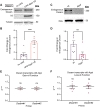
Appl modulates Dscam protein, but not mRNA, expression. (A) Western blot staining for endogenous Dscam (250 kDa), Appl::V5 (150 kDa) and α-Tubulin (50 kDa, as an internal control for normalization) in the central nervous system (CNS) of larvae overexpressing Appl::V5 (UAS-Appl::V5) in all neurons (with nSyb>Gal4) or not (no UAS). (B) Quantification of A shows significant increase in endogenous Dscam expression with pan-neuronal (nSyb>Gal4) overexpression of UAS-Appl::V5 compared to a ‘no UAS’ control (w;;). Two-tailed Mann–Whitney U-test, n=7 groups of 20 larval CNS per genotype, P=0.0023. (C) Western blot staining for endogenous Dscam (250 kDa) and Elav (50 kDa, as an internal control for normalization) in larval CNS that was either wild type or with loss of Appl. (D) Quantification of C shows a significant decrease in endogenous Dscam expression with loss of Appl (Appld) compared to a wild-type control (w;;). Two-tailed Mann–Whitney U-test, n=6 groups of 20 larval VNCs per genotype, P=0.0070. (E,F) Quantification of fold change in endogenous Dscam mRNAs from larval CNS. The data were from RT-qPCR. No change was detected in endogenous Dscam transcript expression with gain (E) or loss (F) of Appl. Transcript expression was normalized internally to Chmp1 (a reference gene) (Kim et al., 2013). Two separate primers (DscamQ3 and DscamQ5) were used to catch all known Dscam isoforms. For each genetic manipulation and primer, significance was assessed. (E) 2−ΔΔCt of Dscam transcripts for pan-neuronal overexpression of Appl (nSyb>Appl::v5) was then divided by the 2−ΔΔCt control (nSyb>mCD8::GFP) to determine the fold change in transcripts. Two-tailed one-sample t-test, n=4 biological replicates per genotype, P=0.9493 (left) and P=0.9998 (right). (F) 2−ΔΔCt of Dscam transcripts for total loss of Appl (Appld) was then divided by the 2−ΔΔCt control (w;;) to determine the fold change in transcripts. Two-tailed one-sample t-test, n=6 biological replicates per genotype, P=0.2132 (left) and P=0.1998 (right). ns, not significant (P>0.05), **P<0.01, ***P<0.001.
Fig. 4.
Fig. 4.
Appl generally affects transmembrane protein expression through a secretion- and intracellular-domain-independent mechanism. (A) Schematic of the domains in full-length Appl and the APP/Appl mutants used in B and C. E1, extracellular domain 1; E2, extracellular domain 2. (B) Axon terminals of all C4da neurons labeled by RFP fluorescence with (1) ppk>no overexpression (wild type; w;;), (2) ppk>UAS-APP695::myc and (3) ppk>UAS-APP695ΔCT::myc. The orange boxes show the magnified segments to the right for viewing the axon connectives, which were individually counted. (C) Average number of axon tracts describes the average of the total number of visible axon connectives between segments A4-A6. Quantification of B shows a significant increase in the number of axon connectives with C4da-specific overexpression of APP695 (UAS-APP695::myc) and APP695 lacking the cytoplasmic domain (UAS-APP695DCT::myc) compared to the control. Kruskal–Wallis with Dunn's Multiple comparisons post hoc analysis, n=9-10 per genotype. (D) Schematic of the domains in the Appl mutants used in E and F. The colors indicate the domains as labeled in panel A. (E) Total C4da neuropil labeled by tdTomato fluorescence with (1) ppk>no overexpression (wild type; w;;), (2) ppk>UAS-ApplΔE2 and (3) ppk>UAS-ApplΔE1. The blue boxes show the magnified segments to the right for viewing the axon connectives, which were individually counted. (F) Quantification of E shows a significant increase in the number of axon connectives when ApplΔE2, but not ApplΔE1, is expressed in C4da neurons. One-way ANOVA with Dunnett's multiple comparisons post hoc analysis. (G) Western blots of endogenous Dscam (250 kDa) and Elav (50 kDa) in the CNS of larvae overexpressing no UAS-Appl transgene (no UAS), UAS-Appl.sdΔE2 or UAS-Appl.sdΔE1 in all neurons (with nSyb>Gal4). (H) Quantification shows that Appl overexpression increases endogenous Dscam expression in the absence of the E2 domain and secretase binding sites, but not in the absence of the E1 domain. Two-tailed one-sample t-test, P=0.0096 (left) and P=0.0186 (right). ns, not significant (P>0.05), *P<0.05, ***P<0.001, ****P<0.0001.
Fig. 5.
Fig. 5.
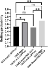
The effects of Appl and Dscam overexpression on nocifensive behavioral response. The graph shows the probability of heat-elicited nocifensive rolling in four genotypes of larvae: (1) UAS-LacZ::GFP.nls (n=145 larvae), (2) UAS-Appl and UAS-LacZ::GFP.nls (n=163 larvae), (3) UAS-DscamTM2::GFP and UAS-LacZ::GFP.nls (n=118 larvae), and (4) UAS-Appl and UAS-DscamTM2::GFP (n=145 larvae). Chi-square tests. ns, not significant (P>0.05), *P<0.05, **P<0.01.

References

    1. Baumann, J. (2007). Down syndrome cell adhesion molecule--a common determinant of brain and heart wiring. Pediatr. Res. 62, 1. 10.1203/PDR.0b013e31811ead83 - DOI - PubMed
    1. Brand, A. H. and Perrimon, N. (1993). Targeted gene expression as a means of altering cell fates and generating dominant phenotypes. Development 118, 401-415. 10.1242/dev.118.2.401 - DOI - PubMed
    1. Bruce, F. M., Brown, S., Smith, J. N., Fuerst, P. G. and Erskine, L. (2017). DSCAM promotes axon fasciculation and growth in the developing optic pathway. Proc. Natl. Acad. Sci. U.S.A. 114, 1702-1707. 10.1073/pnas.1618606114 - DOI - PMC - PubMed
    1. Campbell, G., Weaver, T. and Tomlinson, A. (1993). Axis specification in the developing Drosophila appendage: the role of wingless, decapentaplegic, and the homeobox gene aristaless. Cell 74, 1113-1123. 10.1016/0092-8674(93)90732-6 - DOI - PubMed
    1. Canfield, M. A., Honein, M. A., Yuskiv, N., Xing, J., Mai, C. T., Collins, J. S., Devine, O., Petrini, J., Ramadhani, T. A., Hobbs, C. A.et al. (2006). National estimates and race/ethnic-specific variation of selected birth defects in the United States, 1999-2001. Birth Defects Res. A Clin. Mol. Teratol. 76, 747-756. 10.1002/bdra.20294 - DOI - PubMed

Publication types