Review
. 2023 Sep 15;133(18):e172058.
doi: 10.1172/JCI172058.

Beyond epithelial damage: vascular and endothelial contributions to idiopathic pulmonary fibrosis

Review

Beyond epithelial damage: vascular and endothelial contributions to idiopathic pulmonary fibrosis

James May et al. J Clin Invest. .

Abstract

Idiopathic pulmonary fibrosis (IPF) is a progressive scarring disease of the lung with poor survival. The incidence and mortality of IPF are rising, but treatment remains limited. Currently, two drugs can slow the scarring process but often at the expense of intolerable side effects, and without substantially changing overall survival. A better understanding of mechanisms underlying IPF is likely to lead to improved therapies. The current paradigm proposes that repetitive alveolar epithelial injury from noxious stimuli in a genetically primed individual is followed by abnormal wound healing, including aberrant activity of extracellular matrix-secreting cells, with resultant tissue fibrosis and parenchymal damage. However, this may underplay the importance of the vascular contribution to fibrogenesis. The lungs receive 100% of the cardiac output, and vascular abnormalities in IPF include (a) heterogeneous vessel formation throughout fibrotic lung, including the development of abnormal dilated vessels and anastomoses; (b) abnormal spatially distributed populations of endothelial cells (ECs); (c) dysregulation of endothelial protective pathways such as prostacyclin signaling; and (d) an increased frequency of common vascular and metabolic comorbidities. Here, we propose that vascular and EC abnormalities are both causal and consequential in the pathobiology of IPF and that fuller evaluation of dysregulated pathways may lead to effective therapies and a cure for this devastating disease.


Conflict of interest statement

Conflict of interest: RGJ has received funding from AstraZeneca, Biogen, Galecto, GlaxoSmithKline, Nordic Biosciences, RedX, and Pliant and consulting fees from AstraZeneca, Brainomix, Bristol-Myers Squibb, Chiesi, Cohbar, Daewoong, GlaxoSmithKline, Veracyte, Resolution Therapeutics, and Pliant. RGJ has also received payments from Boehringer Ingelheim, Chiesi, Roche, PatientMPower, AstraZeneca, and Pinsent Masons LLP. RGJ has served on the safety monitoring boards or advisory boards of Boehringer Ingelheim, Galapagos, and Vicore; and served in a leadership role of NuMedii and Action for Pulmonary Fibrosis.

Figures

Figure 1
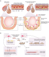
Figure 1. Vascular abnormalities in IPF are reflected within cells, tissue, and the entire organism.
(A) Pulmonary-bronchial anastomoses often develop in IPF and can be visualized by radiography. In healthy tissue, vessels communicate through the capillary bed, and there is an absence of these larger, tortuous communications (23). (B) Fibroblastic foci from fibrotic parenchyma lack ECs (as indicated by an absence of staining for CD34), confirming a lack of vascularity. In healthy tissue, ECs line the vessel walls and are distributed throughout lung tissue. Reproduced with permission from the American Thoracic Society (159). (C) Vascular comorbidities associate with IPF, suggesting that ECs contribute to and are affected by fibrosis.
Figure 2
Figure 2. ECs support healthy vasculature and undergo dramatic changes in IPF.
(A) Damaged epithelium releases active TGF-β and other profibrotic mediators. The original injury also disrupts the BM and the neighboring endothelial layer, which responds to the profibrotic signal. ECs subsequently secrete similar profibrotic mediators and lose the ability to synthesize protective hormones such as eNOS and prostacyclin. This process can stimulate VEGF production, which drives EC proliferation, and ECs distributed throughout the lung propagate fibrosis. Compared with healthy lungs, IPF lungs have a higher proportion of apoptotic ECs, fibroblasts, pericytes, and VSMCs. Cellular proliferation and newly generated vessels expand affected lung tissue. With progressive vascular pathology there is ultimately advanced tissue destruction, and eventually vascular regression develops in the fibroblastic foci. (B) In IPF, the EC participates in several cell-cell interactions and cell transitions. Damaged ECs produce factors that signal to other ECs and promote damage or drive the transition to other cell types: (i) EC-fibroblast: ECs transition into a fibroblast-type cell via EndMT to contribute to the pool of profibrotic cells. (ii) EC-myofibroblast: Damaged ECs also secrete TGF-β, PDGF, and Jag1 to enhance fibroblast-myofibroblast transition and ECM secretion. (iii) EC-EC: Abnormal ECs secrete VEGF, which promotes EC proliferation and abnormal vessel formation, thus contributing to the pool of ECs that can propagate this process. Compromised tight junctions leak coagulation factors, driving fibrosis. (iv) EC-VSMC: EC production of TGF-β and ET1 promotes VSMC proliferation, contributing to PH and a switch to a synthetic phenotype. (v) EC–epithelial cell: Downregulation of protective factors such as MMP-14 delays epithelial repair, allowing persistent epithelial-mesenchymal crosstalk. (vi) Pericyte-myofibroblast: Disrupted Wnt signaling associated with ECs drives pericytes to transition into a myofibroblast-type cell.
Figure 3
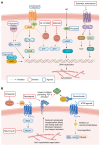
Figure 3. Vascular signaling pathways regulate fibrosis via GPCR, NO, intracellular (PPAR) receptors, and surface integrins in IPF.
(A) Drugs that may counter fibrosis can act through signaling pathways in a range of vascular cell types, including ECs, VSMCs, and fibroblasts. Fibrogenesis-promoting pathways involve GPCRs or NO and signal through cAMP or cGMP to induce fibrosis-related transcriptional events. Treprostinil (TP) acts on cell surface GPCRs to increase intracellular cAMP, which can affect transcription of actin-encoding genes that affect the cytoskeleton, cell motility, and adhesion. TP can also directly activate intracellular PPAR receptors to modulate gene expression. PDE inhibitors (BI-1015550 and sildenafil) prevent cAMP and cGMP breakdown. cGMP, generated following exposure to endogenous NO, activates PKG, which affects gene transcription, the cytoskeleton, and cell contraction. Stimulators, including riociguat, can also generate cGMP. The ET antagonists bosentan and ambrisentan block GPCRs to reduce intracellular Ca2+ concentrations and PKC activity, again modulating gene expression. (B) Therapeutics in IPF can signal through pathways affecting TGF-β signaling or other mechanisms promoting profibrotic gene expression. Ziritaxestat blocks autotaxin, from which LPA is generated. LPA induces various profibrotic effects via GPCRs, including increased RhoA activity and actin cytoskeleton rearrangements that promote altered cell motility in a range of cells relevant to fibrosis. Belumosudil preferentially blocks the ROCK2 isoform. The cytoskeleton can activate cell surface integrins, which are implicated in TGF-β activation. Integrins can be directly blocked by bexotegrast. CTGF, which has numerous profibrotic signaling effects, can be neutralized by the monoclonal antibody pamrevlumab. ATR2 agonists affect numerous intracellular phosphatases, which affect downstream profibrotic gene expression.

Similar articles

Cited by

References

    1. Raghu G, et al. Treatment of idiopathic pulmonary fibrosis with ambrisentan: a parallel, randomized trial. Ann Intern Med. 2013;158(9):641–649. doi: 10.7326/0003-4819-158-9-201305070-00003. - DOI - PubMed
    1. Lederer DJ, Martinez FJ. Idiopathic pulmonary fibrosis. N Engl J Med. 2018;378(19):1811–1823. doi: 10.1056/NEJMra1705751. - DOI - PubMed
    1. Maher TM, et al. Global incidence and prevalence of idiopathic pulmonary fibrosis. Respir Res. 2021;22(1):197. doi: 10.1186/s12931-021-01791-z. - DOI - PMC - PubMed
    1. Hutchinson J, et al. Global incidence and mortality of idiopathic pulmonary fibrosis: a systematic review. Eur Respir J. 2015;46(3):795–806. doi: 10.1183/09031936.00185114. - DOI - PubMed
    1. Strongman H, et al. Incidence, prevalence, and survival of patients with idiopathic pulmonary fibrosis in the UK. Adv Ther. 2018;35(5):724–736. doi: 10.1007/s12325-018-0693-1. - DOI - PMC - PubMed

Publication types