. 2023 Sep 15;133(18):e169199.
doi: 10.1172/JCI169199.

Maternal PlGF and umbilical Dopplers predict pregnancy outcomes at diagnosis of early-onset fetal growth restriction

Affiliations

Maternal PlGF and umbilical Dopplers predict pregnancy outcomes at diagnosis of early-onset fetal growth restriction

Rebecca Spencer et al. J Clin Invest. .

Abstract

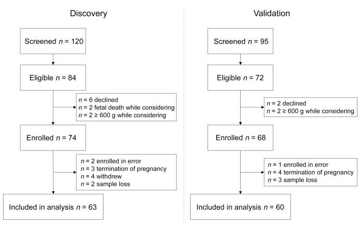
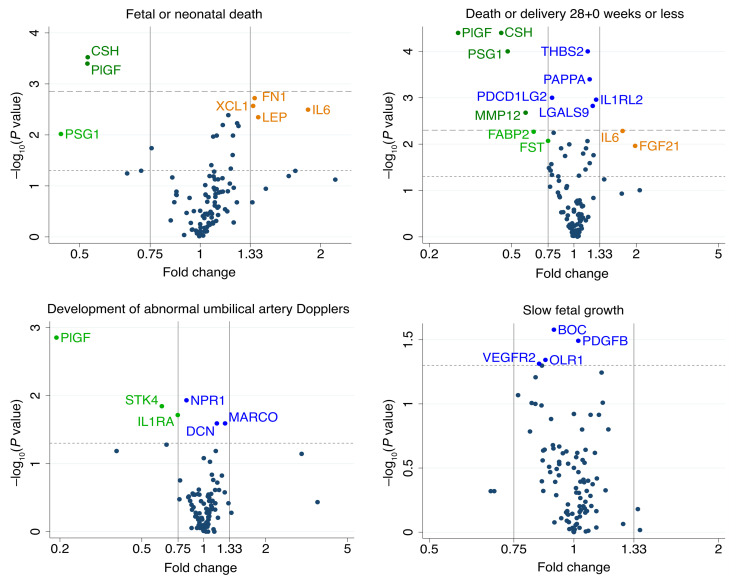
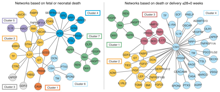
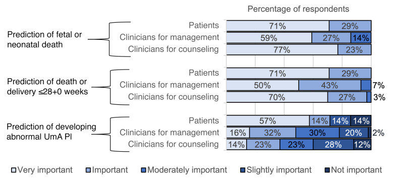
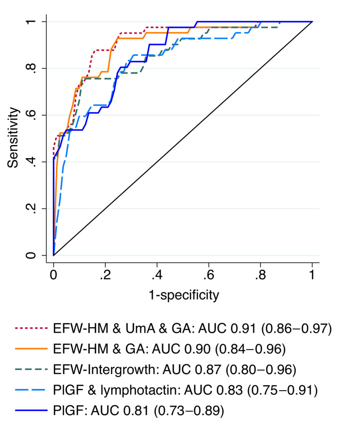
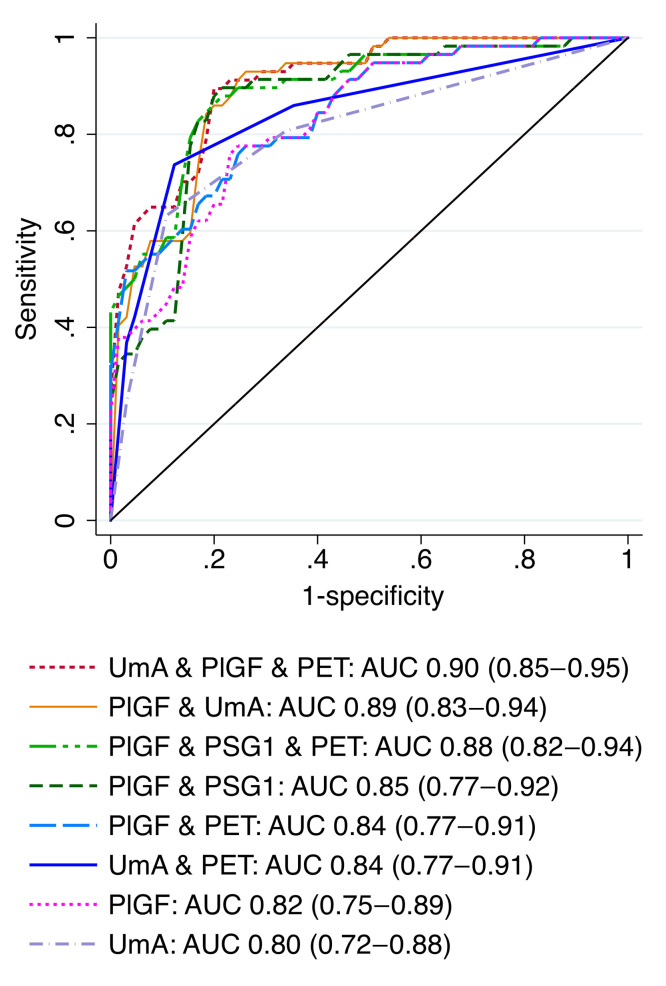
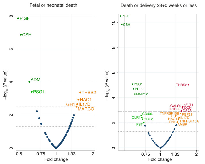
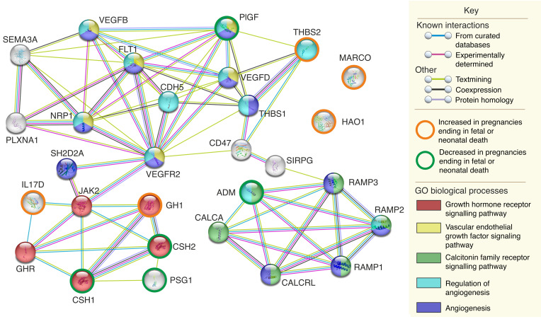
BACKGROUNDSevere, early-onset fetal growth restriction (FGR) causes significant fetal and neonatal mortality and morbidity. Predicting the outcome of affected pregnancies at the time of diagnosis is difficult, thus preventing accurate patient counseling. We investigated the use of maternal serum protein and ultrasound measurements at diagnosis to predict fetal or neonatal death and 3 secondary outcomes: fetal death or delivery at or before 28+0 weeks, development of abnormal umbilical artery (UmA) Doppler velocimetry, and slow fetal growth.METHODSWomen with singleton pregnancies (n = 142, estimated fetal weights [EFWs] below the third centile, less than 600 g, 20+0 to 26+6 weeks of gestation, no known chromosomal, genetic, or major structural abnormalities) were recruited from 4 European centers. Maternal serum from the discovery set (n = 63) was analyzed for 7 proteins linked to angiogenesis, 90 additional proteins associated with cardiovascular disease, and 5 proteins identified through pooled liquid chromatography and tandem mass spectrometry. Patient and clinician stakeholder priorities were used to select models tested in the validation set (n = 60), with final models calculated from combined data.RESULTSThe most discriminative model for fetal or neonatal death included the EFW z score (Hadlock 3 formula/Marsal chart), gestational age, and UmA Doppler category (AUC, 0.91; 95% CI, 0.86-0.97) but was less well calibrated than the model containing only the EFW z score (Hadlock 3/Marsal). The most discriminative model for fetal death or delivery at or before 28+0 weeks included maternal serum placental growth factor (PlGF) concentration and UmA Doppler category (AUC, 0.89; 95% CI, 0.83-0.94).CONCLUSIONUltrasound measurements and maternal serum PlGF concentration at diagnosis of severe, early-onset FGR predicted pregnancy outcomes of importance to patients and clinicians.TRIAL REGISTRATIONClinicalTrials.gov NCT02097667.FUNDINGThe European Union, Rosetrees Trust, Mitchell Charitable Trust.

Keywords: Clinical practice; Diagnostics; Obstetrics/gynecology; Reproductive Biology.


Figures

Figure 1
Figure 1. Flow diagram of participant eligibility and enrollment across the 4 EVERREST Prospective Study centers from March 10, 2014, to January 30, 2020, for the discovery and validation sets.
Figure 2
Figure 2. Volcano plots showing the statistical significance and magnitude of the associations between the 98 proteins and 4 pregnancy outcomes in the discovery set.
Associations were tested with a 2-sided t test for symmetrical data and a Mann-Whitney U test for skewed data. Dotted line indicates a P value of 0.05. Dashed line indicates the Benjamini-Hochberg cutoff with a 5% FDR (P = 0.0015 for fetal or neonatal death, P = 0.005 for death or delivery ≤28+0 weeks). See Supplemental Table 5 for full protein names.
Figure 3
Figure 3. Parenclitic network analysis and clustering by pregnancy outcome.
Networks were generated on the basis of pregnancies ending in fetal or neonatal death versus live births surviving to 29 days of life and pregnancies ending in fetal death or delivery at or before 28+0 weeks of gestation versus continuation of pregnancy beyond 28+0 weeks. See Supplemental Table 5 for full protein names and Supplemental Figures 2 and 3 for the associated dendrograms.
Figure 4
Figure 4. The perceived importance to patients and clinicians of our 3 pregnancy outcomes for model validation.
Importance ranked on a 5-point Likert scale with clinicians asked to separately judge the importance for pregnancy management and the importance for patient counselling.
Figure 5
Figure 5. Comparison of the ROC curves for the models predicting fetal or neonatal death.
EFW-HM, estimated fetal weight calculated using the Hadlock 3 formula (67), with the z score calculated using the Marsal reference chart (68); EFW-Intergrowth, estimated fetal weight and z score calculated using the Intergrowth formula and reference chart (69); GA, gestational age at enrollment; PlGF, placental growth factor concentration. UmA Doppler category: 0 = PI ≤95th centile, 1 = PI >95th centile, 2 = absent EDF, 3 = reversed EDF.
Figure 6
Figure 6. Comparison of the ROC curves for the models predicting fetal death or delivery at or before 28+0 weeks’ gestation.
PET, preeclampsia at enrollment; PSG1 NPX. UmA Doppler category: 0 = PI ≤95th centile, 1 = PI >95th centile, 2 = absent EDF; 3 = reversed EDF.
Figure 7
Figure 7. Volcano plots showing the statistical significance and magnitude of associations between pregnancy outcome and the centered and scaled concentrations of the 93 proteins from the discovery and validation sets combined.
Associations were tested with 2-sided t tests. The dotted line indicates a P value of 0.05; the short-dashed line indicates the Benjamini-Hochberg cutoff with a 5% FDR (P = 0.0048 for fetal or neonatal death, P = 0.012 for death or delivery ≤28+0 weeks); and the long-dashed line indicates the Benjamini-Hochberg cutoff with a 1% FDR (P = 0.00032 for fetal or neonatal death, P = 0.0013 for death or delivery ≤28+0 weeks). See Supplemental Table 10 for full protein names and individual –log10 P values.
Figure 8
Figure 8. An expanded functional network demonstrating interactions and shared GO biological processes of the proteins associated with fetal or neonatal death in the combined data set at a Benjamini-Hochberg FDR of 5%.
See Supplemental Table 5 for full protein names. The analysis, graphics, and legend are from the Swiss Institute of Bioinformatics (STRING) (81).
Figure 9
Figure 9. Functional interactions and shared GO biological processes of the proteins associated with death or delivery at or before 28+0 weeks in the combined data set at a Benjamini-Hochberg FDR of 5%.
See Supplemental Table 5 for full protein names. The analysis, graphics, and legend are from STRING (81).
Figure 10
Figure 10. Observed versus predicted value graphs for models predicting the timing of live birth or fetal death.
Model predicting either the GA of a live-born neonate or the diagnosis of fetal death includes PlGF and sFLT1 concentrations, decorin and matrix metalloproteinase 12 NPX, and UmA Doppler category. Model predicting the interval from enrollment to either live birth or the diagnosis of fetal death includes PlGF and sFLT1 concentrations, decorin and matrix metalloproteinase 12 NPX, UmA Doppler category, and GA at enrollment. Solid green circles indicate pregnancies ending in a live birth; red hollow circles indicate pregnancies ending in fetal death; dotted lines indicate 95% prediction intervals.

Comment in

Similar articles

Cited by

References

    1. Zur RL, et al. The placental basis of fetal growth restriction. Obstet Gynecol Clin North Am. 2020;47(1):81–98. doi: 10.1016/j.ogc.2019.10.008. - DOI - PubMed
    1. Sun C, et al. The placenta in fetal growth restriction: What is going wrong? Placenta. 2020;96:10–18. doi: 10.1016/j.placenta.2020.05.003. - DOI - PubMed
    1. Gordijn SJ, et al. Consensus definition for placental fetal growth restriction: a Delphi procedure. Ultrasound Obstet Gynecol. 2016;48(3):333–339. doi: 10.1002/uog.15884. - DOI - PubMed
    1. Lees CC, et al. ISUOG Practice Guidelines: diagnosis and management of small-for-gestational-age fetus and fetal growth restriction. Ultrasound Obstet Gynecol. 2020;56(2):298–312. doi: 10.1002/uog.22134. - DOI - PubMed
    1. Society for Maternal-Fetal Medicine. et al. Society for Maternal-Fetal Medicine Consult Series #52: Diagnosis and management of fetal growth restriction. Am J Obstet Gynecol. 2020;223(4):B2–B17. doi: 10.1016/j.ajog.2020.05.010. - DOI - PubMed

Publication types

Associated data