. 2023 Sep 15;11(5):e0201423.
doi: 10.1128/spectrum.02014-23. Online ahead of print.

Control of Helicobacter pylori with engineered probiotics secreting selective guided antimicrobial peptides

Affiliations

Control of Helicobacter pylori with engineered probiotics secreting selective guided antimicrobial peptides

Ankan Choudhury et al. Microbiol Spectr. .

Abstract

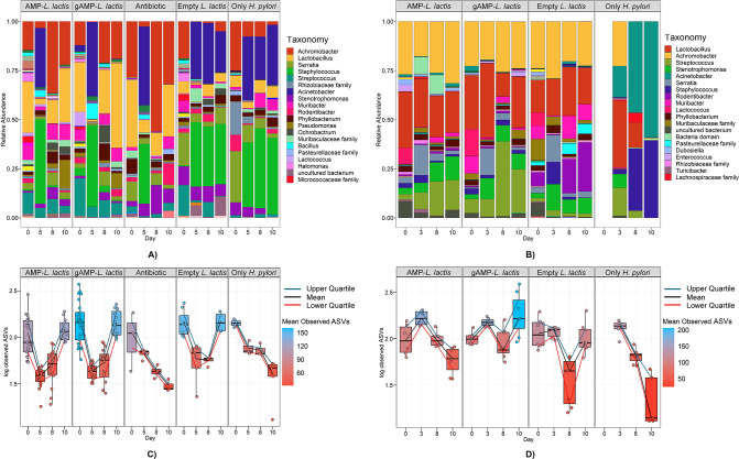
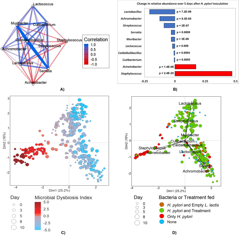
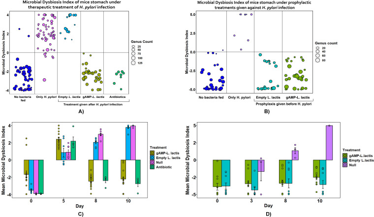
Helicobacter pylori is the primary cause of 78% of gastric cancer cases, providing an opportunity to prevent cancer by controlling a single bacterial pathogen within the complex gastric microbiota. We developed highly selective antimicrobial agents against H. pylori by fusing an H. pylori-binding guide peptide (MM1) to broad-spectrum antimicrobial peptides. The common dairy probiotic Lactococcus lactis was then engineered to secrete these guided antimicrobial peptides (gAMPs). When co-cultured in vitro with H. pylori, the gAMP probiotics lost no toxicity compared to unguided AMP probiotics against the target, H. pylori, while losing >90% of their toxicity against two tested off-target bacteria. To test binding to H. pylori, the MM1 guide was fused to green fluorescent protein (GFP), resulting in enhanced binding compared to unguided GFP as measured by flow cytometry. In contrast, MM1-GFP showed no increased binding over GFP against five different off-target bacteria. These highly selective gAMP probiotics were then tested by oral gavage in mice infected with H. pylori. As a therapy, the probiotics outperformed antibiotic treatment, effectively eliminating H. pylori in just 5 days, and also protected mice from challenge infection as a prophylactic. As expected, the gAMP probiotics were as toxic against H. pylori as the unguided AMP probiotics. However, a strong rebound in gastric species diversity was found with both the selective gAMP probiotics and the non-selective AMP probiotics. Eliminating the extreme microbial dysbiosis caused by H. pylori appeared to be the major factor in diversity recovery. IMPORTANCE Alternatives to antibiotics in the control of Helicobacter pylori and the prevention of gastric cancer are needed. The high prevalence of H. pylori in the human population, the induction of microbial dysbiosis by antibiotics, and increasing antibiotic resistance call for a more sustainable approach. By selectively eliminating the pathogen and retaining the commensal community, H. pylori control may be achieved without adverse health outcomes. Antibiotics are typically used as a therapeutic post-infection, but a more targeted, less disruptive approach could be used as a long-term prophylactic against H. pylori or, by extension, against other gastrointestinal pathogens. Furthermore, the modular nature of the guided antimicrobial peptide (gAMP) technology allows for the substitution of different guides for different pathogens and the use of a cocktail of gAMPs to avoid the development of pathogen resistance.

Keywords: Helicobacter pylori; antimicrobial; dysbiosis; microbiome; mouse; probiotic; selective.


Conflict of interest statement

The authors declare no conflict of interest.

Figures

Fig 1
Fig 1
Precision targeting of H. pylori using probiotic delivery of guided antimicrobial peptides (gAMPs). The probiotic Lactococcus lactis carries the Escherichia coli/L. lactis shuttle vector, pTKR, for the expression of gAMPs in the mouse stomach, allowing rapid engineering in E. coli and transfer to L. lactis. The gAMPs were placed downstream of the acid-inducible P1 promoter and the usp secretion signal peptide to allow secretion in the acidic stomach environment. The guide attached to the N-terminus of the AMP was that portion of the human thrombin protein, Multimerin-1, that binds to H. pylori.
Fig 2
Fig 2
MM1-guided GFP (MM1-GFP) protein binds specifically to H. pylori cells. (A) Protein preparations of MM1-GFP, but not GFP, bound to H. pylori cells. Fluorescence intensity of cells of target bacterium H. pylori 60,190 WT untreated (red) or treated with GFP (green) or MM1-GFP (blue) with averaged median fluorescence (n = 3) in relative fluorescence units (RFUs) obtained from BD FACSverse flow cytometer using blue 488nm laser and a 488/10 bandpass filter; standard deviation shown; statistical significance (one-way analysis of variance, one-way ANOVA; ns, not significant; ***P ≤ 0.001). (B and C) Neither MM1-GFP nor GFP protein significantly bound to off-target bacterial cells. Flow cytometry as in (A) for the cells of off-target bacteria Lactobacillus plantarum (B) or Escherichia coli K12 (C). (D) Confocal microscopy demonstrated that MM1-GFP, but not GFP, bound strongly to Helicobacter pylori cells. Imaging of H. pylori 60,190 WT cells untreated (first column) or treated with GFP (second column) or MM1-GFP (third column). Top row: visualization of GFP or MM1-GFP fluorescence at 488 nm. Middle row: visualization of bacterial cells (CellBrite stain, 640 nm). Bottom row: merged images.
Fig 3
Fig 3
gAMP probiotics selectively kill H. pylori when co-cultured in vitro. Guided (red) or unmodified (blue) versions of three AMPs (alyteserin, CRAMP, and laterosporulin) were expressed by engineered L. lactis, which was co-cultured with the target (H. pylori) or the non-target bacteria, E. coli and Lactobacillus. Eight different initial probiotic concentrations were tested for each AMP or gAMP (x-axis), and the titer of the H. pylori or off-target bacterium was measured after 24 hours of co-culture (y-axis). Titers were determined starting with qPCR using vacA primers for H. pylori, DE3-T7 polymerase primers for E. coli, recA primers for Lactobacillus, and acma primers for L. lactis. The corresponding CFU values were calculated from standard curves of CT versus CFU using CFU values obtained by bacterial dilution and plating (Fig. S3). The limits of qPCR detection differed between bacterial species resulting in flat-lining at different low-end levels.
Fig 4
Fig 4
gAMP and AMP probiotics control H. pylori in the mouse model both as a therapeutic and a prophylactic. (A) For both therapeutic and prophylactic experiments, the H. pylori infection was established by oral gavage for four consecutive days with 250 µL of resuspended H. pylori (~5 × 10⁷ CFU/mL). For therapeutic experiments, the infection was followed by a dose of 250 µL of resuspended L. lactis (~5 × 10⁷ CFU/mL) on day 5 of the regimen. For prophylactic experiments, the probiotic was provided on day 0, followed by a H. pylori challenge on days 3–6. Immediately before administration of both H. pylori and L. lactis, mice stomach samples were extracted by reverse oral gavage method. Further samples were extracted on day 8 and 10 by the same method. For each treatment listed in (B) and (C), at least six mice were used. (B) H. pylori titer measured over the time course of the therapeutic experiment. On each day, probiotics expressing gAMP or AMP were compared to antibiotic treatment or negative controls. H. pylori titers in the reverse oral gavage stomach samples were determined by qPCR using the CFU vs CT standard curve for H. pylori (Fig. S3). The strength of infection is color-coded. Complete data with significance values are presented in Table S2 and S3. (C) H. pylori titer measured over the time course of the prophylactic experiment. The same probiotic and negative control treatments and H. pylori titer determinations were used as in (B).
Fig 5
Fig 5
Probiotic gAMP/AMP treatment in vivo reverses the degradation of taxonomic richness caused by H. pylori infection. (A and C) The left two panels cover the therapeutic experiment, displaying the taxonomic analyses of the samples from Fig. 3B. (B and D) The right two panels cover the prophylactic experiment, with taxonomic analyses of the samples of Fig. 3C. (A and B) The top panels display the major genera found on each sampling day for each treatment, with H. pylori infection leading to domination by Acinetobacter and Staphylococcus and a relief from this domination being provided by the probiotic treatments. (C and D) The bottom panels chart the taxonomic richness by a simple ASV log10 count over time for each treatment group, with a rebound or retention of taxonomic richness in the probiotic treatment group, especially the gAMP probiotic group.
Fig 6
Fig 6
The Microbial Dysbiosis Index (MDI) comprised 10 co-varying taxa that also correlated with H. pylori infection. (A) Compositional Correlation network of 10 genera revealed by the CCREPE analysis, whose composition co-varied among the mice stomach samples on days 0 and 5, which were pre- and post-H. pylori infection, respectively. Eight of these were positively correlated with each other (blue lines), while two others (Staphylococcus and Acinetobacter) were positively correlated with each other but inversely correlated with the other 8 (red lines). All the correlations were significant (P < 0.05, q < 0.10). (B) The 10 genera that co-varied significantly also had a significant change in their mean relative abundance among the samples of day 0 vs day 5, with 8 of them decreasing following H. pylori infection while Staphylococcus and Acinetobacter saw a significant increase. This further validates the importance of these genera as markers for mice stomach microbial health in our experiment. (C) Principal Components Analysis (PCA) of the taxonomic relationship between the microbiota of all 350 samples in the study. Dysbiotic samples (red) clustered separately from non-dysbiotic samples (blue), demonstrating that MDI correlated with the taxonomic relatedness of the samples generally. Jitter was used to allow all samples to be in view. (D) The same PCA as in (C) but with ordinates overlayed corresponding to the 10 genera of the MDI, with longer vectors indicating more correspondence to the abundance of that genus. Staphylococcus and Acinetobacter are again seen as strong predictors for H. pylori/no treatment samples (red). No jitter was used in order to report the unaltered PCA output.
Fig 7
Fig 7
Probiotic gAMP treatment protects against microbial dysbiosis. (A) At the start of the therapeutic experiment (day 0), the MDIs of all samples (dark blue) were negative (healthy) and had predominantly high taxonomic richness (circle size). After H. pylori infection and probiotic treatment (days 8 and 10 combined), untreated (“only H. pylori”) and empty probiotic-treated samples were dysbiotic with low taxonomic richness, while AMP and gAMP probiotic-treated samples were healthy and had high taxonomic richness. Antibiotic treatment relieved dysbiosis but had low taxonomic richness. (B) In the prophylactic experiment, all probiotic treatments protected against H. pylori-induced dysbiosis, with gAMP probiotics promoting the most robust taxonomic richness. Infected mice without the prophylactic yielded only dysbiotic samples. (C) Dysbiosis in the therapeutic experiment, tracked by day. All treatments showed a negative (healthy) MDI for day 0 and a positive (dysbiotic) MDI for day 5 since these samples were collected just before oral inoculation with H. pylori or probiotic, respectively. The bar colors correspond to the colors and treatments in (A). Within 3 days, dysbiosis had been alleviated by gAMP and AMP probiotics and antibiotics but not by the empty vector probiotic. (D) Dysbiosis in the prophylactic experiment, tracked by day. All samples had healthy MDI values before (day 0) and 3 days after probiotic treatment, but H. pylori induced dysbiosis by days 8 and 10 in samples lacking prophylaxis (null treatment, magenta). All probiotic treatments protected against H. pylori-induced dysbiosis.

References

    1. Khatun S, Appidi T, Rengan AK. 2021. The role played by bacterial infections in the onset and metastasis of cancer. Curr Res Microb Sci 2:100078. doi: 10.1016/j.crmicr.2021.100078 - DOI - PMC - PubMed
    1. van Elsland D, Neefjes J. 2018. Bacterial infections and cancer. EMBO Rep 19:e46632. doi: 10.15252/embr.201846632 - DOI - PMC - PubMed
    1. Clay SL, Fonseca-Pereira D, Garrett WS. 2022. Colorectal cancer: the facts in the case of the microbiota. J Clin Invest 132:e155101. doi: 10.1172/JCI155101 - DOI - PMC - PubMed
    1. International agency for research on cancer . 2012. A review of human carcinogens - PubMed
    1. Yang L, Kartsonaki C, Yao P, de Martel C, Plummer M, Chapman D, Guo Y, Clark S, Walters RG, Chen Y, Pei P, Lv J, Yu C, Jeske R, Waterboer T, Clifford GM, Franceschi S, Peto R, Hill M, Li L, Millwood IY, Chen Z, China Kadoorie Biobank Collaborative Group . 2021. The relative and attributable risks of cardia and non-cardia gastric cancer associated with Helicobacter pylori infection in China: a case-cohort study. Lancet Public Health 6:e888–e896. doi: 10.1016/S2468-2667(21)00164-X - DOI - PMC - PubMed

LinkOut - more resources