. 2023 Apr-Jun;35(2):117-146.
doi: 10.5935/2965-2774.20230310-en.

The use of bedside echocardiography in the care of critically ill patients - a joint consensus document of the Associação de Medicina Intensiva Brasileira, Associação Brasileira de Medicina de Emergência and Sociedade Brasileira de Medicina Hospitalar. Part 2 - Technical aspects

[Article in English, Portuguese]
Affiliations

The use of bedside echocardiography in the care of critically ill patients - a joint consensus document of the Associação de Medicina Intensiva Brasileira, Associação Brasileira de Medicina de Emergência and Sociedade Brasileira de Medicina Hospitalar. Part 2 - Technical aspects

[Article in English, Portuguese]
José Augusto Santos Pellegrini et al. Crit Care Sci. 2023 Apr-Jun.

Abstract

Echocardiography in critically ill patients has become essential in the evaluation of patients in different settings, such as the hospital. However, unlike for other matters related to the care of these patients, there are still no recommendations from national medical societies on the subject. The objective of this document was to organize and make available expert consensus opinions that may help to better incorporate echocardiography in the evaluation of critically ill patients. Thus, the Associação de Medicina Intensiva Brasileira, the Associação Brasileira de Medicina de Emergência, and the Sociedade Brasileira de Medicina Hospitalar formed a group of 17 physicians to formulate questions relevant to the topic and discuss the possibility of consensus for each of them. All questions were prepared using a five-point Likert scale. Consensus was defined a priori as at least 80% of the responses between one and two or between four and five. The consideration of the issues involved two rounds of voting and debate among all participants. The 27 questions prepared make up the present document and are divided into 4 major assessment areas: left ventricular function, right ventricular function, diagnosis of shock, and hemodynamics. At the end of the process, there were 17 positive (agreement) and 3 negative (disagreement) consensuses; another 7 questions remained without consensus. Although areas of uncertainty persist, this document brings together consensus opinions on several issues related to echocardiography in critically ill patients and may enhance its development in the national scenario.


Conflict of interest statement

Conflicts of interest: None.

Figures

Figure 1
Figure 1
Parasternal longitudinal window.
Figure 2
Figure 2
Several observation planes in the transverse parasternal window. (A) Patient in the left lateral decubitus position. Transducer in the third left intercostal space, with the index pointed to the left shoulder (2 hours). (B) Transducer with tip tilted upward to visualize the section at the level of the aortic valve (see asterisk). (C) Less inclined transducer, obtaining a section at the level of the mitral valve (see arrow). (D) Transducer with tip inclined downward, visualizing the section at the level of the papillary muscles (see arrows).
Figure 3
Figure 3
Fourand five-chamber apical windows. (A) Patient in the left lateral semidecubitus position (slightly inclined toward the back). Transducer in the fifth left intercostal space, between the midclavicular line and the anterior axillary line, with the index pointed to the left arm (3 o’clock). (B) Four-chamber apical window. (C) Apical five-chamber window: obtained from the apical four-chamber window, with the tip of the transducer tilted slightly upward, maintaining contact with the patient’s skin, in which the aortic valve and the left ventricular outflow tract can be seen.
Figure 4
Figure 4
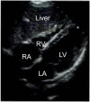
Four-chamber subcostal window, where the liver can also be visualized.
Figure 5
Figure 5
Subcostal window of the inferior vena cava.
Figure 6
Figure 6
Measurement of tissue Doppler S’ wave. (A) Positioning of the tissue Doppler cursor on the lateral wall of the mitral annulus (arrow) in the apical four-chamber view. (B) Tissue Doppler curve in a patient with normal systolic function, in which we can visualize the systolic wave and the E’ and A’ diastolic waves. Peak velocity of the S’ wave with normal amplitude (S’ wave > 9cm/s).
Figure 7
Figure 7
Measurement of the systolic excursion of the tricuspid annulus plane. (A) Positioning of the M-mode cursor at the level of the lateral base of the tricuspid annulus (arrow) in the four-chamber apical window. (B) M-mode waveform depicting the movement of the lateral base of the tricuspid ring during the cardiac cycle. The ascending phase of the tracing corresponds to systole. The systolic excursion of the tricuspid annulus plane is measured as the height of the wave. In this patient, the systolic excursion of the tricuspid annulus plane was 22.8mm (normal value > 17mm).
Figure 8
Figure 8
Tissue Doppler imaging of the peak velocity of tricuspid annulus displacement during right ventricular systole (tissue S’ wave).
Figure 9
Figure 9
Estimated maximum velocity of tricuspid regurgitation (approximately 3m/sec). First, we must locate the jet with color Doppler imaging. Next, we align the Doppler cursor (dashed line) with the jet and select the continuous Doppler function. Then, in the speed record, a continuous curve appears.
Figure 10
Figure 10
Tissue Doppler ultrasound of the basal lateral wall of the left ventricle. Note the E’ wave below the baseline during diastole (E’ or e’ wave).

Similar articles

Cited by

References

    1. Vieillard-Baron A, Millington SJ, Sanfilippo F, Chew M, Diaz-Gomez J, McLean A, et al. A decade of progress in critical care echocardiography: a narrative review. Intensive Care Med. 2019;45(6):770–788. - PubMed
    1. Mayo PH, Beaulieu Y, Doelken P, Feller-Kopman D, Harrod C, Kaplan A, et al. American College of Chest Physicians/La Societe de Reanimation de Langue Francaise statement on competence in critical care ultrasonography. Chest. 2009;135(4):1050–1060. - PubMed
    1. Expert Round Table on Echocardiography in ICU International consensus statement on training standards for advanced critical care echocardiography. Intensive Care Med. 2014;40(5):654–666. - PubMed
    1. Levitov A, Frankel HL, Blaivas M, Kirkpatrick AW, Su E, Evans D, et al. Guidelines for the Appropriate Use of Bedside General and Cardiac Ultrasonography in the Evaluation of Critically Ill Patients-Part II: Cardiac Ultrasonography. Crit Care Med. 2016;44(6):1206–1227. - PubMed
    1. Pellegrini JA, Cordioli RL, Grumann AC, Ziegelmann PK, Taniguchi LU. Point-of-care ultrasonography in Brazilian intensive care units: a national survey. Ann Intensive Care. 2018;8(1):50. - PMC - PubMed