. 2023 Sep 15;27(3):26.
doi: 10.1007/s00792-023-01309-z.

Structure/function studies of the NAD+-dependent DNA ligase from the poly-extremophile Deinococcus radiodurans reveal importance of the BRCT domain for DNA binding

Affiliations

Structure/function studies of the NAD+-dependent DNA ligase from the poly-extremophile Deinococcus radiodurans reveal importance of the BRCT domain for DNA binding

Andreia Fernandes et al. Extremophiles. .

Abstract

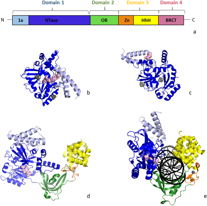
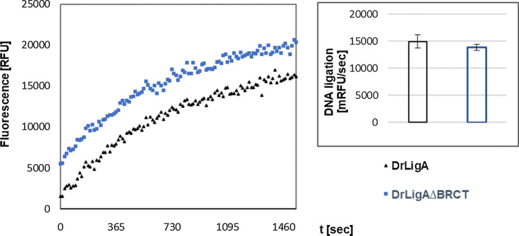
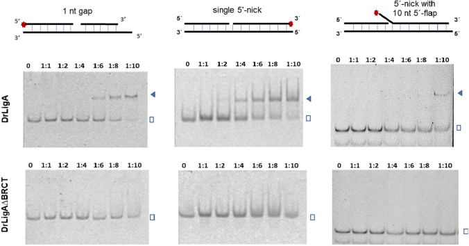
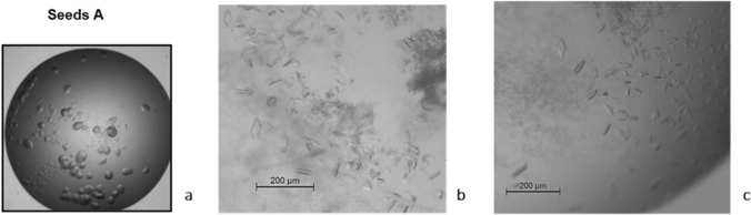
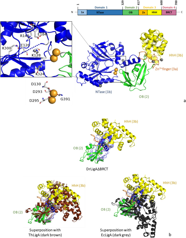
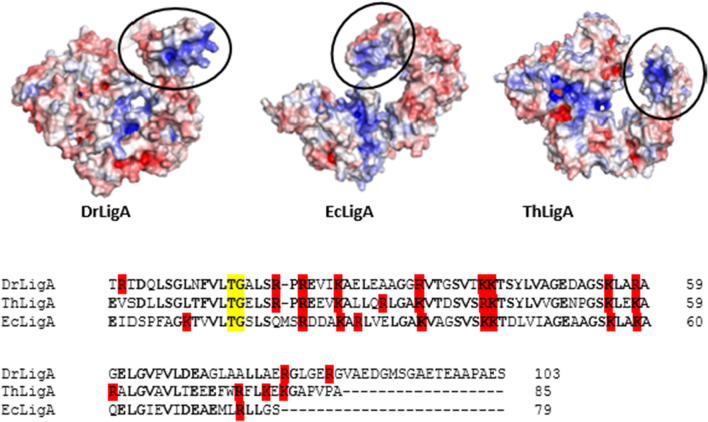
Bacterial NAD+-dependent DNA ligases (LigAs) are enzymes involved in replication, recombination, and DNA-repair processes by catalyzing the formation of phosphodiester bonds in the backbone of DNA. These multidomain proteins exhibit four modular domains, that are highly conserved across species, with the BRCT (breast cancer type 1 C-terminus) domain on the C-terminus of the enzyme. In this study, we expressed and purified both recombinant full-length and a C-terminally truncated LigA from Deinococcus radiodurans (DrLigA and DrLigA∆BRCT) and characterized them using biochemical and X-ray crystallography techniques. Using seeds of DrLigA spherulites, we obtained ≤ 100 µm plate crystals of DrLigA∆BRCT. The crystal structure of the truncated protein was obtained at 3.4 Å resolution, revealing DrLigA∆BRCT in a non-adenylated state. Using molecular beacon-based activity assays, we demonstrated that DNA ligation via nick sealing remains unaffected in the truncated DrLigA∆BRCT. However, DNA-binding assays revealed a reduction in the affinity of DrLigA∆BRCT for dsDNA. Thus, we conclude that the flexible BRCT domain, while not critical for DNA nick-joining, plays a role in the DNA binding process, which may be a conserved function of the BRCT domain in LigA-type DNA ligases.

Keywords: BRCT; DNA ligase A; DNA nick-joining; Protein–DNA binding; X-ray crystallography.


Figures

Fig. 1
Fig. 1
Representation of the modular organization and structures of bacterial DNA ligases A a identification of the domains and subdomains: Domain 1 with subdomain 1a (light blue) and 1b/NTase (blue), Domain 2/OB (green), Domain 3 with subdomain 3a/Zn (orange) and 3b/HhH (yellow), and Domain 4/BRCT (purple). NTase nucleotidyl transferase, OB oligonucleotide-binding, Zn zinc finger, HhH helix–hairpin–helix, BRCT breast cancer type 1 C-terminus. b E. faecalis truncated LigA (PDB ID: 1tae). c E. faecalis truncated LigA (PDB: 1ta8), d T. filiformis apo-LigA (PDB ID: 1dgs). e E. coli LigA–DNA complex (PDB ID: 2owo). Metals (gray) and cofactor (light pink) are shown as spheres, and DNA is colored black
Fig. 2
Fig. 2
Thermofluor results of DrLigA: non normalized graphs and melting temperatures of the most stabilizing buffer conditions. *Reference buffer representing buffer 1
Fig. 3
Fig. 3
Molecular beacon-based activity assays with 330 nM of DrLigA: DNA ligation by nick closure. a Representation of the molecular beacon with a single 5′-nick which is phosphorylated at the 5′-end and contains the fluorophore–quencher pair TAMRA-Dabcyl. b and c The increase in fluorescence was measured as relative fluorescence units (RFU) over time, and the DNA ligation activity is represented as the initial velocity, which data were normalized to 100%. b pH effect: dotted line—buffer 20 mM Tris–HCl 50 mM NaCl, solid line—20 mM Bis–Tris 50 mM NaCl. c Salt concentration effect: experiment performed with 5 μM NAD+ in buffer 20 mM Bis–Tris pH 7.0, 50 mM NaCl. Experiments were performed in duplicate. Error bars represent 1 standard deviation from the mean (1 S.D.)
Fig. 4
Fig. 4
Molecular beacon-based activity assays with 170 nM of DrLigA: NAD+ concentration influence on nick closure. The increase in fluorescence was measured as relative fluorescence units (RFU) over time, and the DNA ligation activity is represented as the initial velocity, which data were normalized to 100%. Experiments were performed with 20 mM buffer Bis–Tris pH 6.5 and 50 mM of NaCl. Error bars represent 1 standard deviation from the mean (1 S.D.)
Fig. 5
Fig. 5
Molecular beacon-based activity assay with 66 nM of protein: DNA ligation by nick closure with DrLigA full-length versus truncated DrLigA∆BRCT. The increase in fluorescence was measured as relative fluorescence units (RFU) over time, and the ligation activity is represented as the initial velocity in mRFU per sec. Experiments performed at pH 7.0, with 1 mM Mn2+ and 5 µM NAD+. Curves represent the mean values (duplicates). Column graph error bars represent 1 standard deviation from the mean (1 S.D.)
Fig. 6
Fig. 6
Gel-shift assays with 100 nM of FAM-labelled DNA substrates: DNA binding of DrligA full-length versus truncated DrLigA∆BRCT. The incubation of DNA substrates (with the 5′-end of the internal nick/gap not phosphorylated) and proteins was performed for 15 min at room temperature and an increasing concentration of protein was added. 0, 1:1, 1:2, 1:4, 1:6, 1:8, and 1:10 represent the molar ratio of [DNA]: [protein]. The blue triangles and the open squares indicate the DNA–protein complex and free DNA, respectively
Fig. 7
Fig. 7
DrLigA and DrLigA∆BRCT crystallization results: a DrLigA spherulites obtained in solution A (1 M sodium acetate trihydrate, 0.1 M sodium HEPES pH 7.5, 0.05 M cadmium sulfate 8/3 hydrate) by sitting drop vapor diffusion. They were used for seeding (seeds A). b and c Crystals of DrLigA∆BRCT obtained by microseeding with seeds A. b DrLigA∆BRCT crystals grew in solution B (20% w/v PEG 8000, 0.2 M magnesium acetate tetrahydrate, and 0.1 M sodium cacodylate pH 6.5). c DrLigA∆BRCT crystals obtained in solution B with 0.01 M trimethylamine hydrochloride as additive
Fig. 8
Fig. 8
Structure of DrLigA∆BRCT: a modular organization of DrLigA and overall structure of DrLigA∆BRCT with NTase subdomain 1b (blue), OB-fold domain 2 (green), zinc finger motif 3a (orange), and HhH subdomain 3b (yellow) identified. On the left, amplified view of the two-metal center and catalytic site. K128 from KXDG motif, residues E126, R149, E184, K300, and K324 (predicted to interact with NAD+) (top view), and the residues D130, D293, D295, and G391 (predicted to interact with Mn2+) (bottom view) are labelled. Mn2+ cations are represented as light orange spheres, and Zn2+ cations as light gray spheres. b Representation of superimposed DrLigA∆BRCT (alignment of subdomain 1b) with ThLigA structure (PDB ID: 1dgs, dark brown) and with EcLigA structure (PDB ID: 5tt5, dark gray). NTase nucleotidyl transferase, OB oligonucleotide-binding, Zn zinc finger, HhH helix–hairpin–helix, BRCT breast cancer type 1 C-terminus
Fig. 9
Fig. 9
Electrostatic surface (blue positive charges, red negative charges) from bacterial DNA ligases A structures predicted by AlphaFold2—DrLigA (D. radiodurans, UniProtKB: Q9RSQ5), EcLigA (E. coli, UniProtKB: P15042), ThLigA (T. filiformis, UniProtKB: Q9ZHI0). Black ellipse identifies the BRCT domain. Multiple sequence alignment from Clustal Omega (https://www.ebi.ac.uk/Tools/msa/clustalo/), alignment of BRCT domain from DrLigA, ThLigA, and EcLigA. Positively charged residues Arg and Lys are highlighted in red. Conserved residues from the phosphate-binding pocket are colored yellow

Similar articles

References

    1. Blasius M, Buob R, Shevelev IV, Hubscher U. Enzymes involved in DNA ligation and end-healing in the radioresistant bacterium Deinococcus radiodurans. BMC Mol Biol. 2007;8:69. doi: 10.1186/1471-2199-8-69. - DOI - PMC - PubMed
    1. Burley SK, Berman HM, Bhikadiya C, et al. Protein data bank: the single global archive for 3D macromolecular structure data. Nucleic Acids Res. 2019;47:D520–D528. doi: 10.1093/nar/gky949. - DOI - PMC - PubMed
    1. Callebaut I, Mornon JP. From BRCA1 to RAP1: a widespread BRCT module closely associated with DNA repair. FEBS Lett. 1997;400:25–30. doi: 10.1016/S0014-5793(96)01312-9. - DOI - PubMed
    1. Clapperton JA, Manke IA, Lowery DM, et al. Structure and mechanism of BRCA1 BRCT domain recognition of phosphorylated BACH1 with implications for cancer. Nat Struct Mol Biol. 2004;11:512–518. doi: 10.1038/nsmb775. - DOI - PubMed
    1. Cowtan K. The Buccaneer software for automated model building. 1. Tracing protein chains. Acta Crystallogr D Biol Crystallogr. 2006;62:1002–1011. doi: 10.1107/S0907444906022116. - DOI - PubMed

LinkOut - more resources