. 2024 Feb;42(2):190-200.
doi: 10.1007/s11604-023-01487-y. Epub 2023 Sep 15.

Preliminary assessment of automated radiology report generation with generative pre-trained transformers: comparing results to radiologist-generated reports

Affiliations

Preliminary assessment of automated radiology report generation with generative pre-trained transformers: comparing results to radiologist-generated reports

Takeshi Nakaura et al. Jpn J Radiol. 2024 Feb.

Abstract

Purpose: In this preliminary study, we aimed to evaluate the potential of the generative pre-trained transformer (GPT) series for generating radiology reports from concise imaging findings and compare its performance with radiologist-generated reports.

Methods: This retrospective study involved 28 patients who underwent computed tomography (CT) scans and had a diagnosed disease with typical imaging findings. Radiology reports were generated using GPT-2, GPT-3.5, and GPT-4 based on the patient's age, gender, disease site, and imaging findings. We calculated the top-1, top-5 accuracy, and mean average precision (MAP) of differential diagnoses for GPT-2, GPT-3.5, GPT-4, and radiologists. Two board-certified radiologists evaluated the grammar and readability, image findings, impression, differential diagnosis, and overall quality of all reports using a 4-point scale.

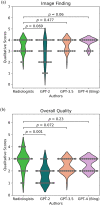
Results: Top-1 and Top-5 accuracies for the different diagnoses were highest for radiologists, followed by GPT-4, GPT-3.5, and GPT-2, in that order (Top-1: 1.00, 0.54, 0.54, and 0.21, respectively; Top-5: 1.00, 0.96, 0.89, and 0.54, respectively). There were no significant differences in qualitative scores about grammar and readability, image findings, and overall quality between radiologists and GPT-3.5 or GPT-4 (p > 0.05). However, qualitative scores of the GPT series in impression and differential diagnosis scores were significantly lower than those of radiologists (p < 0.05).

Conclusions: Our preliminary study suggests that GPT-3.5 and GPT-4 have the possibility to generate radiology reports with high readability and reasonable image findings from very short keywords; however, concerns persist regarding the accuracy of impressions and differential diagnoses, thereby requiring verification by radiologists.

Keywords: Computed tomography; Deep learning; Generative pre-trained transformer; Large language model; Radiology report.


Conflict of interest statement

Toshinori Hirai has received research support from Canon Medical Systems.

Figures
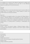
Fig. 1
Fig. 1
An example of “Prompt” and “Information of a patient”. Before inputting actual patient data into the GPT series, it is necessary to provide guidance on the role and type of text to be generated. This instruction is called a “Prompt”. The prompt serves as a way to inform the language model about the context and desired output. In this case, the example prompt explains that the output should be from the perspective of a radiologist and the purpose of each part of the radiology report. The “Prompt” is common for all patients, and only the “Main text” portion varies for each individual patient. This approach ensures that the language model receives consistent contextual information while tailoring the generated report to the specific details of each patient's case
Fig. 2
Fig. 2
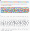
A visualization of tokens used in generative pre-trained transformer (GPT) series. GPT series and other transformer-based models perform language processing (a) in units called “tokens”, each of which has a unique identifier (b). The task of text generation is internally processed as selecting the token with the highest probability of appearing after a particular sequence of tokens. This approach allows the model to generate coherent and contextually appropriate text by predicting and selecting the most likely tokens to follow a given input sequence
Fig. 3
Fig. 3
Qualitative analysis. Violin plots show qualitative analysis of the image findings (a) and the overall quality (b)
Fig. 4
Fig. 4
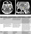
A 69-year-old female patient with a suspected pituitary adenoma. Non-contrast CT axial image (a), contrast-enhanced CT sagittal image (b) and generated radiology reports by GPT series (c) are shown. A tumor with homogeneous enhancement is observed from the sella turcica to the suprasellar region, suggesting a pituitary adenoma. Information input other than the prompt is “Age (years): 69, sex: female, modality: contrast enhanced CT, location: the vicinity of the sella turcica, diameter: 49 mm, findings: enhancing supra- and intrasellar mass”. The GPT-2 report is a simple report written according to the input information, and the differential diagnosis seems relatively reasonable. In the GPT-3.5 report, both the findings and impression sections are more detailed than in the GPT-2 report. The GPT-4.0 report is overall quite similar to a human-generated report, and the differential diagnosis is reasonable. However, it includes information that was not input, such as calcification and cystic degeneration
Fig. 5
Fig. 5
A 31-year-old female with a hepatic hemangioma. The contrast-enhanced CT arterial phase (a) shows heterogeneous enhancement within the lesion, and the venous phase (b) reveals a generally stronger enhancement than the liver parenchyma, consistent with typical findings of a hemangioma. Generated radiology reports by GPT series (c) are also shown. The information inputted besides the prompt is “Age (years): 31; sex: female; modality: contrast enhanced CT; location: segment 8 in the liver; diameter: 22 mm; findings: delayed phase-enhancing lesion”. In the GPT-2 generated report, a list of differential diagnoses is not even created, and the impression primarily suspects hepatocellular carcinoma. In the GPT-3.5 generated report, although the format is well organized, hepatocellular carcinoma is still listed as the top differential diagnosis. The GPT-4.0 generated report is generally quite good, with reasonable differential diagnoses
Fig. 6
Fig. 6
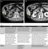
A 75-year-old male with an angiomyolipoma in the right kidney. A non-contrast CT (a) and contrast-enhanced CT (b) reveal a fatty renal mass in the right kidney. Generated radiology reports by GPT series (c) are also shown. The information inputted besides the prompt is “Age (years): 75; sex: male; modality: contrast-enhanced CT; location: the inferior portion of the right kidney; diameter: 35 mm; findings: fat-containing renal mass”. In the GPT-2 generated report, the possibility of a renal tumor accompanied by surrounding edema is low, and a list of differential diagnoses is not even created. In the GPT-3.5 generated report, although the lesion is located in the kidney, the differential diagnoses include adrenal adenoma. In the GPT-4.0 generated report, the overall quality is quite good; however, there is a description of “calcification” in the image findings, which was not part of the input information

Similar articles

Cited by

References

    1. Hartung MP, Bickle IC, Gaillard F, Kanne JP. How to create a great radiology report. Radiographics. 2020;40:1658–1670. doi: 10.1148/rg.2020200020. - DOI - PubMed
    1. Parikh JR, Wolfman D, Bender CE, Arleo E. Radiologist burnout according to surveyed radiology practice leaders. J Am Coll Radiol. 2020;17:78–81. doi: 10.1016/j.jacr.2019.07.008. - DOI - PubMed
    1. Kitahara H, Nagatani Y, Otani H, Nakayama R, Kida Y, Sonoda A, et al. A novel strategy to develop deep learning for image super-resolution using original ultra-high-resolution computed tomography images of lung as training dataset. Jpn J Radiol. 2022;40:38–47. doi: 10.1007/s11604-021-01184-8. - DOI - PMC - PubMed
    1. Barat M, Chassagnon G, Dohan A, Gaujoux S, Coriat R, Hoeffel C, et al. Artificial intelligence: a critical review of current applications in pancreatic imaging. Jpn J Radiol. 2021;39:514–523. doi: 10.1007/s11604-021-01098-5. - DOI - PubMed
    1. Chassagnon G, De Margerie-Mellon C, Vakalopoulou M, Marini R, Hoang-Thi T-N, Revel M-P, et al. Artificial intelligence in lung cancer: current applications and perspectives. Jpn J Radiol. 2023;41:235–244. - PMC - PubMed