. 2023 Oct;10(30):e2301730.
doi: 10.1002/advs.202301730. Epub 2023 Sep 15.

Living Material with Temperature-Dependent Light Absorption

Affiliations

Living Material with Temperature-Dependent Light Absorption

Lealia L Xiong et al. Adv Sci (Weinh). 2023 Oct.

Abstract

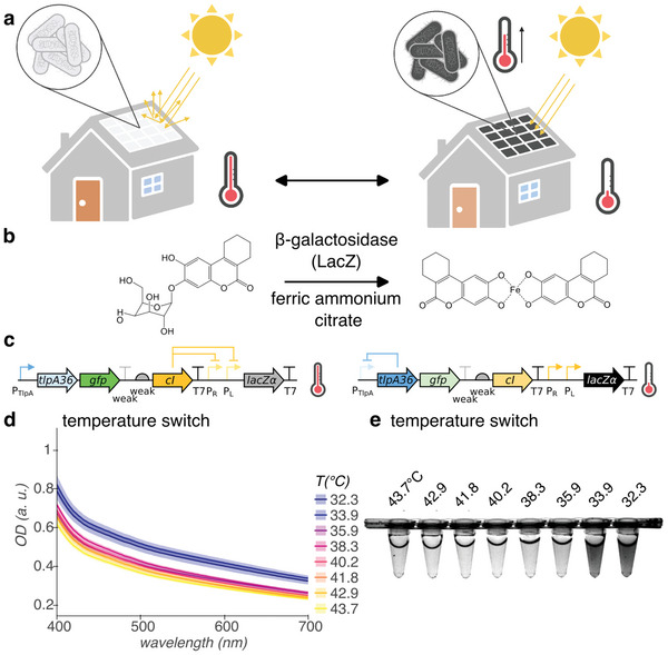
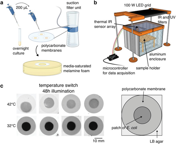
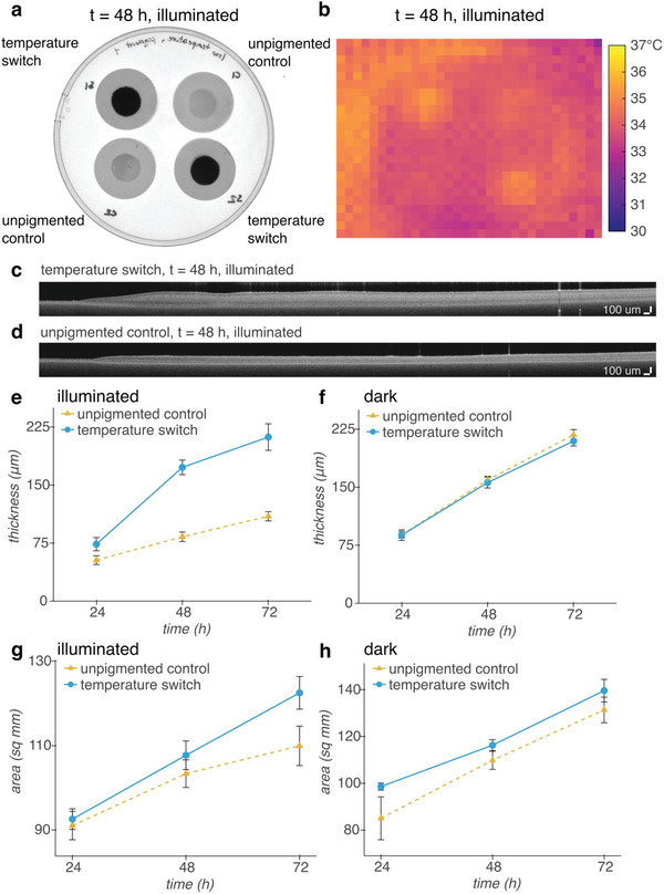
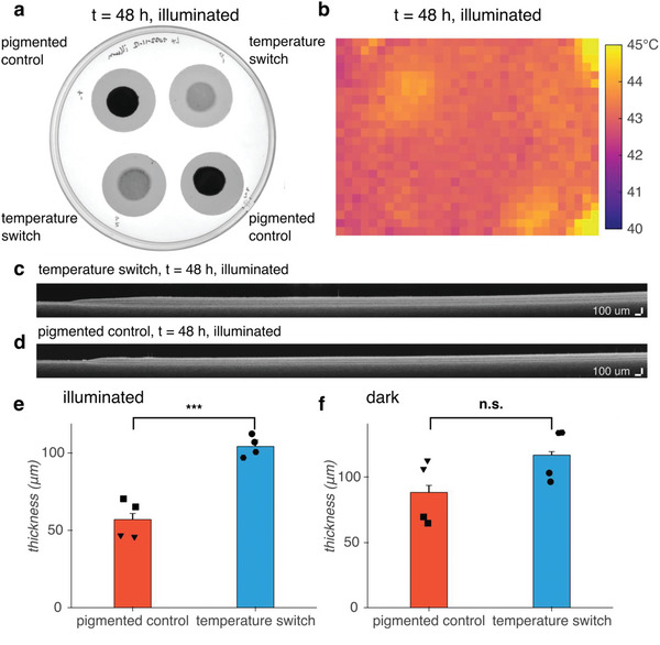
Engineered living materials (ELMs) exhibit desirable characteristics of the living component, including growth and repair, and responsiveness to external stimuli. Escherichia coli (E. coli) are a promising constituent of ELMs because they are very tractable to genetic engineering, produce heterologous proteins readily, and grow exponentially. However, seasonal variation in ambient temperature presents a challenge in deploying ELMs outside of a laboratory environment because E. coli growth rate is impaired both below and above 37 °C. Here, a genetic circuit is developed that controls the expression of a light-absorptive chromophore in response to changes in temperature. It is demonstrated that at temperatures below 36 °C, the engineered E. coli increase in pigmentation, causing an increase in sample temperature and growth rate above non-pigmented counterparts in a model planar ELM. On the other hand, at above 36 °C, they decrease in pigmentation, protecting the growth compared to bacteria with temperature-independent high pigmentation. Integrating the temperature-responsive circuit into an ELM has the potential to improve living material performance by optimizing growth and protein production in the face of seasonal temperature changes.

Keywords: engineered living materials; synthetic biology; thermal control.


Conflict of interest statement

The authors declare no conflict of interest.

Figures

Figure 1
Figure 1
Cold‐activated production of light‐absorptive pigment for E. coli‐containing ELMs. a) Illustration of ELM used as building material. At ambient temperature greater than or equal to optimum for growth, E. coli remains colorless (left). However, at ambient temperatures less than optimal, E. coli express light‐absorptive pigment, warming under illumination by the sun to recover growth rate (right). Illustration created with BioRender.com. b) Genetically‐encodable light‐absorptive pigment system. β‐galactosidase cleaves S‐gal at the glycosidic bond, exposing the esculetin group, which coordinates with ferric iron to form a black pigment. c) Circuit diagram of temperature switch construct for low‐temperature pigmentation, with state of regulation arcs, indicated at high and low temperatures. Genes, left to right: tlpA36, gfp, cI, lacZα. d,e) Visible light optical density (OD) spectra d) and representative white light transillumination image e) of cultures of E. coli containing the temperature switch construct after 24 h growth in pigment‐induction media at temperatures ranging from 43.7 °C to 32.3 °C. n = 4 biological replicates; shading represents +/− standard error of the mean.
Figure 2
Figure 2
Temperature‐dependent pigmentation in a model ELM. a) Schematic of formation of dense, centimeter‐scale patches of E. coli to simulate the ELM environment. We grew E. coli overnight to saturation in a liquid medium. For each patch, we transferred 200 µL of culture to track‐etched polycarbonate membranes (25 mm diam., 0.2 µm pores) and applied suction to coat the cells onto the membranes, forming a dense patch. We then transferred the coated membranes to a melamine foam substrate saturated with liquid media for growth. b) Schematic of illuminated growth chamber. We used a 100 W white light LED to expose E. coli patches to illumination and monitored the temperature using a 32 × 24 array of thermal IR sensors. The sensor array is attached to a motorized arm and retracts when not imaging to avoid shadowing the samples. c) Transillumination white light images of patches of E. coli containing the temperature switch construct on polycarbonate membranes after 48 h growth in the illuminated growth chamber with pigment‐induction media at 42 °C and 32 °C. Parts of figure created with BioRender.com.
Figure 3
Figure 3
Cold‐induced pigment improves the growth of dense patches of E. coli under illumination at 32 °C. a) White light transillumination image of patches of E. coli containing either our temperature switch construct or an unpigmented control construct encoding heat‐inducible GFP after transferring to agar for imaging at 48 h. b) Thermal IR image of patches inside illuminated growth chamber at 48 h. c,d) Representative OCT images of patches of E. coli containing our temperature c) switch construct, or d) the unpigmented control construct. e,f) Thickness of patches grown e) under illumination, or f) in a dark incubator over time. The slower rate of evaporation without illumination allows for thicker growth overall than with illumination. g,h) Area of patches grown g) under illumination or h) in a dark incubator over time. n = 4 biological replicates; error bars represent +/− standard error of the mean.
Figure 4
Figure 4
Turning off pigmentation above 36 °C improves the growth of dense patches of E. coli under illumination at 42 °C compared to a pigmented control. a) White light transillumination image of patches of E. coli containing either the temperature switch construct or a pigmented control construct encoding IPTG‐inducible LacZα after transferring to agar for imaging at 48 h. b) Thermal IR image of patches inside illuminated growth chamber at 48 h. c,d) Representative OCT images of patches of E. coli containing our c) temperature switch construct or d) the pigmented control construct. e,f) Thickness of patches grown e) under illumination (p = 0.0006) or f) in a dark incubator (p = 0.1214) at 48 h. Inverted triangle and hexagon markers indicate patches coated onto Whatman Nucleopore polycarbonate membranes; square and circle markers indicate patches coated onto Sartorius polycarbonate membranes. n = 4 biological replicates; error bars represent +/− standard error of the mean. p‐values calculated using a two‐tailed unpaired t‐test; *** p ≤ 0.001, n.s. p ≥ 0.05.

Similar articles

Cited by

  • Harnessing Fungi Signaling in Living Composites.
    Schyck S, Marchese P, Amani M, Ablonczy M, Spoelstra L, Jones M, Bathaei Y, Bismarck A, Masania K. Schyck S, et al. Glob Chall. 2024 Jul 12;8(8):2400104. doi: 10.1002/gch2.202400104. eCollection 2024 Aug. Glob Chall. 2024. PMID: 39469481 Free PMC article.

References

    1. Nguyen P. Q., Courchesne N.‐M. D., Duraj‐Thatte A., Praveschotinunt P., Joshi N. S., Adv. Mater. 2018, 30, 1704847. - PMC - PubMed
    1. Gilbert C., Ellis T., ACS Synth. Biol. 2019, 8, 1. - PubMed
    1. Liu S., Xu W., Front. Sens. 2020, 1.
    1. Heveran C. M., Williams S. L., Qiu J., Artier J., Hubler M. H., Cook S. M., Cameron J. C., Srubar W. V., Matter 2020, 2, 481.
    1. Rothschild L. J., Biochem. Soc. Trans. 2016, 44, 1158. - PMC - PubMed

Publication types

LinkOut - more resources