. 2023 Sep 26;42(9):113141.
doi: 10.1016/j.celrep.2023.113141. Epub 2023 Sep 19.

Mast cell deficiency improves cognition and enhances disease-associated microglia in 5XFAD mice

Affiliations

Mast cell deficiency improves cognition and enhances disease-associated microglia in 5XFAD mice

Chih-Chung Jerry Lin et al. Cell Rep. .

Abstract

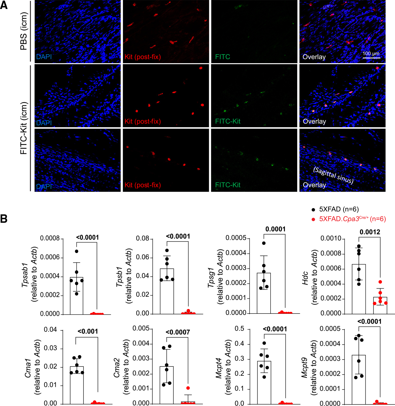
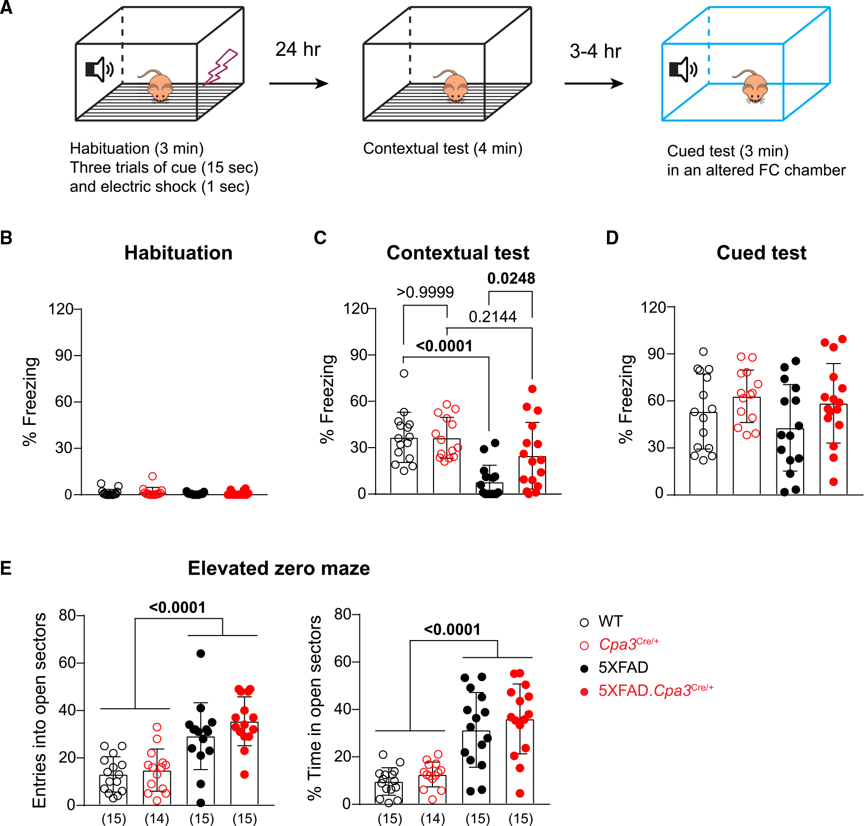
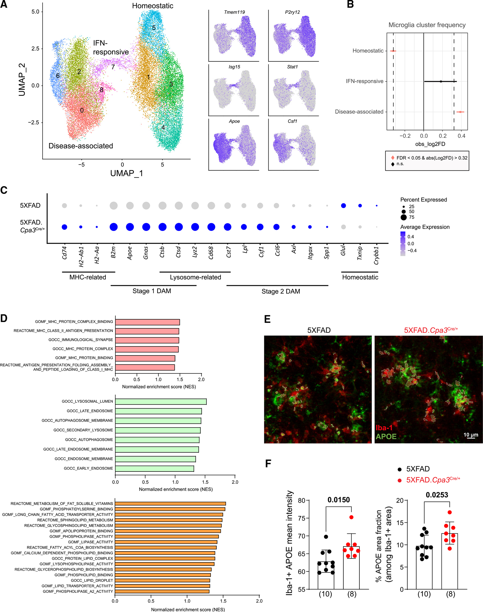
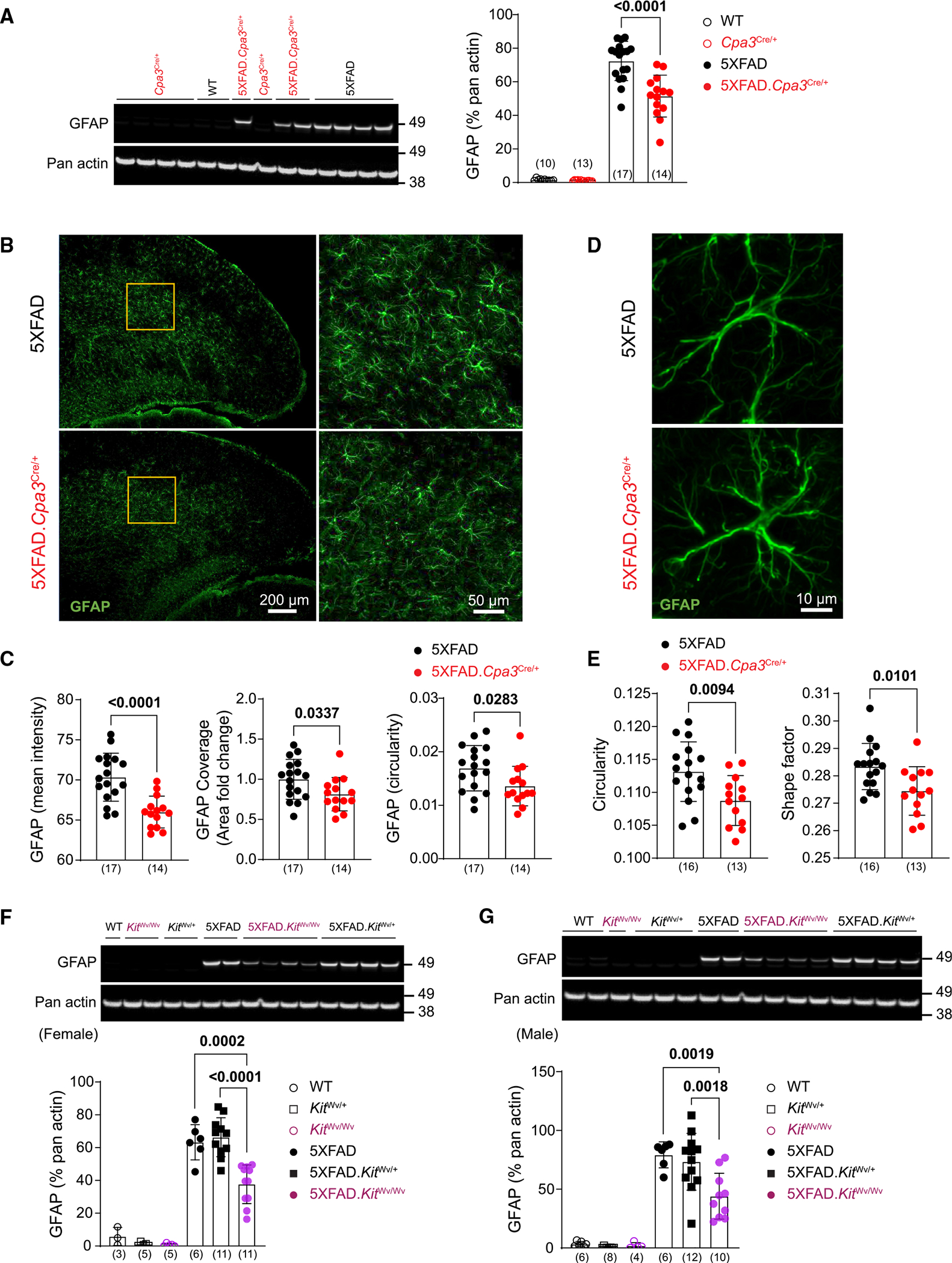
Emerging evidence suggests that peripheral immune cells contribute to Alzheimer's disease (AD) neuropathogenesis. Among these, mast cells are known for their functions in allergic reactions and neuroinflammation; however, little is known about their role in AD. Here, we crossed 5XFAD mice with mast cell-deficient strains and observed the effects on AD-related neuropathology and cognitive impairment. We found that mast cell depletion improved contextual fear conditioning in 5XFAD mice without affecting cued fear conditioning, anxiety-like behavior, or amyloid burden. Furthermore, mast cell depletion led to an upregulation of transcriptomic signatures for putatively protective disease-associated microglia and resulted in reduced markers indicative of reactive astrocytes. We hypothesize a system of bidirectional communication between dural mast cells and the brain, where mast cells respond to signals from the brain environment by expressing immune-regulatory mediators, impacting cognition and glial cell function. These findings highlight mast cells as potential therapeutic targets for AD.

Keywords: 5XFAD; Alzheimer's disease; CP: Immunology; CP: Neuroscience; astrocyte; behavior; mast cell; meninges; microglia; neuroimmune interaction.


Conflict of interest statement

Declaration of interests R.E.T. is a consultant to and shareholder in AZTherapeutics. R.I.S. is a shareholder in Feromics.

Figures

Figure 1.
Figure 1.. Dural mast cells can scan cerebrospinal fluid contents and are required for meningeal expression of genes encoding immune-relevant effector proteins
(A) Immunofluorescence images of dura harvested from mice intracisternally injected with PBS or FITC-labeled anti-KIT antibody. The tissues were then fixed and stained with anti-Kit antibody detected by secondary antibody conjugated with Alexa Fluor 568. Data are from one representative experiment of two (n = 1–2 mice/group). (B) Quantitative PCR analysis of mast cell mediator genes in dura samples harvested from 12- to 13-month-old female 5XFAD and 5XFAD.Cpa3Cre/+ mice (n = 6/group). Bar graphs depict mean ± SD with individual mice as points. Unpaired t tests were performed to determine significance. See also Figure S1.
Figure 2.
Figure 2.. Depletion of mast cells restores contextual fear conditioning without affecting anxiety-like behavior in 5XFAD mice
1-year-old female WT, Cpa3Cre/+, 5XFAD, and 5XFAD.Cpa3Cre/+ mice were evaluated in the fear conditioning (FC) test and elevated zero maze. (A) Illustration of the 2-day FC test procedure. (B–D) The freezing frequency during habituation (B), contextual FC test (C), and cued FC test (D). (E) Number of entries and percent time spent in the open sectors in elevated zero maze. The number of mice is specified in the panels. Bar graphs depict mean ± SD with individual mice as points. Two-way ANOVA with Tukey’s multiple comparison tests were performed to determine significance. See also Figure S2.
Figure 3.
Figure 3.. Depletion of mast cells increases transcriptomic profiles of disease-associated microglia and impacts multiple biological pathways
(A) Brain cells were isolated from the cortical regions of eight 8-month-old female 5XFAD and 5XFAD.Cpa3Cre/+ mice (n = 4/genotype under cold conditions and single-cell sequenced). UMAP plot of 31,351 single-cell RNA microglia profiles were partitioned into nine transcriptional subclusters and grouped into three microglial states based on their feature genes: homeostatic (Tmem119, P2ry12), IFN-responsive (Isg15, Stat1), and disease-associated ones (Apoe, Csf1). (B) Changes in frequency of microglial states in 5XFAD and 5XFAD.Cpa3Cre/+ brains. (C) Dot plot of DAM, MHC, lysosomal, and homeostatic genes of disease-associated state of microglia in (A). Expression level (color scale) of marker genes in two genotypes and the percentage of cells expressing them (dot size). (D) GSEA analysis identified several MHC, endo-lysosome, and lipid pathways that were upregulated in mast cell-deficient disease-associated microglia subclusters compared with mast cell-sufficient microglia (false discovery rate q values < 0.25). (E) Brain cortical sections were labeled with Iba-1 (red) and APOE (green) antibodies. Representative images of the visual area of isocortex from 8- to 9-month-old female 5XFAD (n = 10) and 5XFAD.Cpa3Cre/+ (n = 8) mice. (F) Quantification of mean fluorescence intensity and coverage of APOE among Iba-1-positive microglia. Bar graphs depict mean ± SD with individual mice as points. Unpaired t test was performed to determine significance. IFN, interferon; MHC, major histocompatibility complex; GSEA; gene set enrichment analysis. See also Figure S3 and Table S1.
Figure 4.
Figure 4.. Mast cell deficiency reduces GFAP expression and alters astrocyte morphology in 5XFAD mice
(A) Representative western blots and protein levels for reactive astrocyte marker GFAP measured from cortical homogenates of 8- to 9-month-old female Cpa3Cre cohort mice. (B) Brain cortical sections were labeled with GFAP (green) antibody. Representative images of cortex from 8- to 9-month-old female 5XFAD and 5XFAD.Cpa3Cre/+ mice. The details of GFAP-positive astrocytes in the inserts are shown. (C) Quantification of mean fluorescence intensity, area fold change, and circularity of GFAP-positive cells in (B). (D) Representative images of a single astrocyte in the visual cortex of 8- to 9-month-old female 5XFAD and 5XFAD.Cpa3Cre/+ mice. (E) Quantification of GFAP-positive astrocyte circularity and shape factor in (D). (F and G) Representative western blots and protein levels for astrogliosis marker GFAP measured from cortical homogenates of 7- to 9-month-old female (F) or 8- to 10-month-old male (G) KitWv cohort mice. GFAP, glial fibrillary acidic protein. Bar graphs depict mean ± SD with individual mice as points. The number of mice is specified in the panels. Two-way ANOVA with Tukey’s multiple comparison tests (A, F, and G) and unpaired t test (C and E) were performed to determine significance. See also Figure S4.

Similar articles

Cited by

References

    1. Knopman DS, Amieva H, Petersen RC, Chételat G, Holtzman DM, Hyman BT, Nixon RA, and Jones DT (2021). Alzheimer disease. Nat. Rev. Dis. Primers 7, 33. 10.1038/s41572-021-00269-y. - DOI - PMC - PubMed
    1. Chen X, and Holtzman DM (2022). Emerging roles of innate and adaptive immunity in Alzheimer’s disease. Immunity 55, 2236–2254. 10.1016/J.IMMUNI.202. - DOI - PMC - PubMed
    1. Jorfi M, Maaser-Hecker A, and Tanzi RE (2023). The neuroimmune axis of Alzheimer’s disease. Genome Med. 15, 6. 10.1186/S13073-023-01155-W. - DOI - PMC - PubMed
    1. Jones MK, Nair A, and Gupta M (2019). Mast cells in neurodegenerative disease. Front. Cell. Neurosci. 13, 171–179. 10.3389/fncel.2019.00171. - DOI - PMC - PubMed
    1. Forsythe P (2019). Mast Cells in Neuroimmune Interactions. Trends Neurosci. 42, 43–55. 10.1016/j.tins.2018.09.006. - DOI - PubMed

Publication types

Substances