. 2023 Sep 15;18(9):e0286256.
doi: 10.1371/journal.pone.0286256. eCollection 2023.

Inducible overexpression of a FAM3C/ILEI transgene has pleiotropic effects with shortened life span, liver fibrosis and anemia in mice

Affiliations

Inducible overexpression of a FAM3C/ILEI transgene has pleiotropic effects with shortened life span, liver fibrosis and anemia in mice

Ulrike Schmidt et al. PLoS One. .

Abstract

FAM3C/ILEI is an important factor in epithelial-to-mesenchymal transition (EMT) induction, tumor progression and metastasis. Overexpressed in many cancers, elevated ILEI levels and secretion correlate with poor patient survival. Although ILEI's causative role in invasive tumor growth and metastasis has been demonstrated in several cellular tumor models, there are no available transgenic mice to study these effects in the context of a complex organism. Here, we describe the generation and initial characterization of a Tet-ON inducible Fam3c/ILEI transgenic mouse strain. We find that ubiquitous induction of ILEI overexpression (R26-ILEIind) at weaning age leads to a shortened lifespan, reduced body weight and microcytic hypochromic anemia. The anemia was reversible at a young age within a week upon withdrawal of ILEI induction. Vav1-driven overexpression of the ILEIind transgene in all hematopoietic cells (Vav-ILEIind) did not render mice anemic or lower overall fitness, demonstrating that no intrinsic mechanisms of erythroid development were dysregulated by ILEI and that hematopoietic ILEI hyperfunction did not contribute to death. Reduced serum iron levels of R26-ILEIind mice were indicative for a malfunction in iron uptake or homeostasis. Accordingly, the liver, the main organ of iron metabolism, was severely affected in moribund ILEI overexpressing mice: increased alanine transaminase and aspartate aminotransferase levels indicated liver dysfunction, the liver was reduced in size, showed increased apoptosis, reduced cellular iron content, and had a fibrotic phenotype. These data indicate that high ILEI expression in the liver might reduce hepatoprotection and induce liver fibrosis, which leads to liver dysfunction, disturbed iron metabolism and eventually to death. Overall, we show here that the novel Tet-ON inducible Fam3c/ILEI transgenic mouse strain allows tissue specific timely controlled overexpression of ILEI and thus, will serve as a versatile tool to model the effect of elevated ILEI expression in diverse tissue entities and disease conditions, including cancer.


Conflict of interest statement

The authors have declared that no competing interests exist.

Figures

Fig 1
Fig 1. Generation of KH2-ILEI-FLAG ES cells.
(A) Scheme of the flp-in targeting strategy in KH2 ES cells. Adapted from [30]. (B) FLAG Western blot analysis of KH2 ES cells after transient transfection with the Fam3c/ILEI-FLAG transgene targeting construct with or without Dox induction. Erk1 was used as loading control. (C) Validation of transgene targeted insertion by PCR on single clones of KH2 ES cells after Hygromycin selection. Clone IDs in black were selected for further analysis. (D) FLAG western blot analysis of sub-selected KH2-ILEI-FLAG clones with or without Dox induction. Erk1 was used as loading control. Clone IDs in black were selected for further analysis. (E) Southern blot analysis of sub-selected KH2-ILEI-FLAG clones with a probe specific for the engineered locus. KH2 cells were used as negative and KH2-STAT1 cells [30] as positive controls. Clone #7 in bold was selected for blastocyst injection.
Fig 2
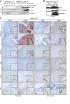
Fig 2. The Fam3c/ILEI-FLAG transgene is broadly expressed in R26-ILEIind mice upon doxycycline induction.
(A) Western blot analysis of bone marrow isolated from control Rosa26rtTA and dual-transgenic Rosa26rtTA-ILEIind (R26-ILEIind) mice after ex vivo culture for 24 or 48 hours in the presence of the indicated Dox concentrations. FLAG and ILEI antibodies were used for transgene detection, beta-actin was used as the loading control. (B) ILEI western blot analysis of spleen, bone marrow, and blood sera freshly isolated from R26-ILEIind mice kept on normal or switched to Dox drinking water (1 mg/ml Dox and 5% sucrose) 3 days before sacrifice. Please note, splenocytes also showed endogenous ILEI expression. (C) ILEI and FLAG immunohistochemistry on thin sections of the intestine, kidney, liver, lung, and skin of R26-ILEIind mice kept on normal or switched to Dox drinking water (1 mg/ml Dox and 5% sucrose) 3 days before sacrifice. Scale bar, 50μm; scale bar for inlets, 20μm.
Fig 3
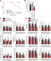
Fig 3. Inducible ILEI overexpression results in reduced life span, body weight, and microcytic hypochromic anemia.
(A) Survival plot of ILEIind and R26-ILEIind mice kept on normal or switched to Dox drinking water at 3 weeks of age. (B) Mean body weight ± SEM of R26-ILEIind mice kept on Dox drinking water measured one week before death (dark data points) or at experiment endpoint (white data points) compared to age-matched mice used as Dox treatment and genetic controls. (C) Mean number of WBC, CD11b+, Gr1+, CD11b and Gr1 double-positive, CD3+ and CD19+ cells ± SEM measured at last three weeks before death of R26-ILEIind mice kept on Dox water and compared to age-matched mice used as Dox treatment and genetic controls. (D) Mean HGB concentration (top left), RBC count (middle left) HCT percentage (bottom left), MCV (top right), MCH (middle right) and MCHC (bottom right) ± SEM measured at last three weeks before death of R26-ILEIind mice kept on Dox water and compared to age-matched mice used as Dox treatment and genetic controls. (B-D) Statistical significance was determined by one-way ANOVA and is marked with asterisks (*p<0.05; **p<0.01; ***p<0.001; ****p<0.0001).
Fig 4
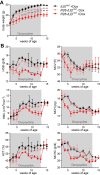
Fig 4. Anemic phenotype is reversible upon withdrawal of temporary ILEI overexpression.
(A, B) Mean (A) body weight and (B) HGB concentration (top left), RBC count (middle left), HCT percentage (bottom left), MCV (top right), MCH (middle right) and MCHC (bottom right) ± SEM of R26-ILEIind mice kept temporarily on Dox water from week 3 to 13 of age and measured weekly until week 15 of age compared to littermates used as Dox treatment and genetic controls. (A, B) Statistical significance was determined by one-way ANOVA and is marked with asterisks (*p<0.05; **p<0.01; ***p<0.001; ****p<0.0001).
Fig 5
Fig 5. ILEI overexpression in hematopoietic cells does not influence overall fitness and red blood cell parameters.
(A) Western blot analysis of freshly isolated bone marrow from dual-transgenic Vav-ILEIind mice kept on Dox supplemented either in the drinking water or in the food at different concentrations for 3 days. FLAG and ILEI antibodies were used for transgene detection, vinculin as loading control. (B) Mean body weight ± SEM of ILEIind and Vav-ILEIind mice kept on normal or Dox diet. (C) Mean percentage of live cells ± SEM of listed myeloid subpopulations in the spleen of ILEIind and Vav-ILEIind mice kept on normal or Dox diet. (D) Mean HGB concentration, RBC count and HCT percentage ± SEM of Vav-ILEIind mice kept temporarily on Dox diet from week 3 to 10 of age and measured weekly until week 12 of age compared to littermates used as Dox treatment and genetic controls. Statistical significance was determined by student’s t-test and is marked with asterisks (*p<0.05; **p<0.01; ***p<0.001).
Fig 6
Fig 6. Ubiquitous ILEI overexpression leads to reduced iron levels in the serum and in hepatocytes and to liver dysfunction.
(A) Mean serum iron levels ± SEM of R26-ILEIind mice kept on Dox drinking water measured one week before death compared to age-matched mice used as Dox treatment and genetic controls. (B-C) Mean (B) ALT and (C) AST levels ± SEM of R26-ILEIind mice kept on Dox drinking water measured one week before death (dark data points) or at experiment endpoint (white data points) compared to age-matched mice used as Dox treatment and genetic controls. (D) Mean liver-to-body weight ratio ± SEM of R26-ILEIind mice kept on Dox drinking water measured one week before death (dark data points) or at experiment endpoint (white data points) compared to age-matched mice used as Dox treatment and genetic controls. (E-F) Representative (E) macroscopic images and (F) H&E stained thin sections of the liver of moribund R26-ILEIind mice kept on Dox drinking water compared to age-matched mice used as Dox treatment and genetic controls. (G-L) Mean percentage of (G) Ki67 and (I) activated Caspase 3 (actCasp3) positive cells ± SEM with representative images of corresponding (H) Ki67 and (J) actCasp3 immunohistochemistry of the liver of moribund R26-ILEIind mice kept on Dox drinking water compared to age-matched mice used as Dox treatment and genetic controls. (K,L) Iron content of the liver shown as (K) Fe3+ positive hepatocytes per mm2 ± SEM and (L) representative images of moribund R26-ILEIind mice kept on Dox drinking water compared to age-matched mice used as Dox treatment and genetic controls. Scale bars, (E) 1 cm, (F,H,J,L) 100μm. Statistical significance was determined by one-way ANOVA (A,B,C,D,G,I) or student’s t-tests (K) and marked with asterisks (*p<0.05; **p<0.01; ***p<0.001; ****p<0.0001).
Fig 7
Fig 7. Mice with induced ubiquitous ILEI overexpression develop liver fibrosis.
(A-D) Representative images of (A) Fibronectin and (B) S100A4 immunohistochemistry and their quantifications shown as mean percentage of (C) Fibronectin and (D) S100A4 positive area ± SEM of the liver of moribund R26-ILEIind mice kept on Dox drinking water compared to age-matched mice used as Dox treatment and genetic controls. (E-F) Increased collagen deposition and bile duct density shown by (E) Sirius Picric Red staining and (F) E-cadherin immunohistochemistry. Scale bar, 200μm; scale bar for inlets, 50μm. (B,C) Statistical significance was determined by one-way ANOVA and marked with asterisks (*p<0.05; ****p<0.0001).

References

    1. Zhu Y, Xu G, Patel A, McLaughlin MM, Silverman C, Knecht K, et al.. Cloning, expression, and initial characterization of a novel cytokine-like gene family. Genomics. 2002;80(2):144–50. doi: 10.1006/geno.2002.6816 - DOI - PubMed
    1. Jechlinger M, Grunert S, Tamir IH, Janda E, Ludemann S, Waerner T, et al.. Expression profiling of epithelial plasticity in tumor progression. Oncogene. 2003;22(46):7155–69. doi: 10.1038/sj.onc.1206887 - DOI - PubMed
    1. Jansson AM, Csiszar A, Maier J, Nystrom AC, Ax E, Johansson P, et al.. The interleukin-like epithelial-mesenchymal transition inducer ILEI exhibits a non-interleukin-like fold and is active as a domain-swapped dimer. The Journal of biological chemistry. 2017;292(37):15501–11. doi: 10.1074/jbc.M117.782904 - DOI - PMC - PubMed
    1. Johansson P, Bernstrom J, Gorman T, Oster L, Backstrom S, Schweikart F, et al.. FAM3B PANDER and FAM3C ILEI represent a distinct class of signaling molecules with a non-cytokine-like fold. Structure. 2013;21(2):306–13. doi: 10.1016/j.str.2012.12.009 - DOI - PubMed
    1. Kral M, Klimek C, Kutay B, Timelthaler G, Lendl T, Neuditschko B, et al.. Covalent dimerization of interleukin-like epithelial-to-mesenchymal transition (EMT) inducer (ILEI) facilitates EMT, invasion, and late aspects of metastasis. The FEBS journal. 2017;284(20):3484–505. doi: 10.1111/febs.14207 - DOI - PubMed

Publication types